Chapter 13 Orbit
INTRODUCTION
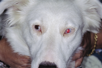
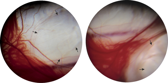
Figure 13-3 Two views of the same eye of the dog shown in Figure 13-2. This Australian shepherd has hereditary multiple ocular anomalies. A large staphyloma (arrows) is present in the peripheral fundus, with choroidal dysplasia (choroidal vessels not formed). The purple streak is the long posterior ciliary artery.
Figure 13-2 Coloboma of the iris associated with hereditary multiple ocular anomalies in an Australian shepherd.
< div class='tao-gold-member'>
Only gold members can continue reading. Log In or Register to continue
Stay updated, free articles. Join our Telegram channel

Full access? Get Clinical Tree


