Chapter 1 Eyelid
INTRODUCTION
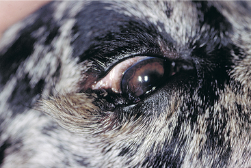
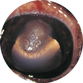
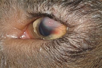
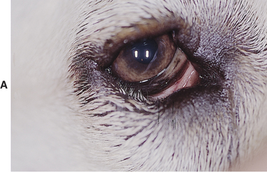
Figure 1-3 Right eye of cat in Figure 1-2. The upper eyelid agenesis involves most of the eyelid and lateral canthus—the most common location for eyelid agenesis in cats. Pigment is present on the cornea as a result of chronic exposure and because hair has come in contact with the cornea. The defect is corrected by the grafting of skin and conjunctiva to fill the defect. A mature cataract is present and is an unrelated problem.
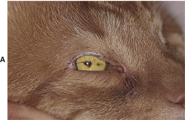
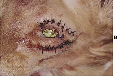
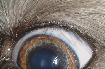
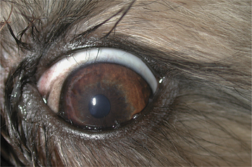
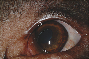
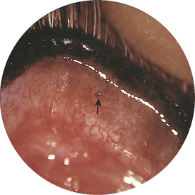
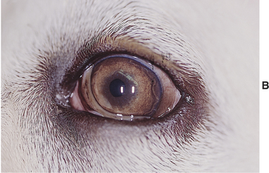
Figure 1-11 Single, stout upper eyelid distichium in the upper eyelid of a 7-year-old Labrador retriever.
< div class='tao-gold-member'>
Only gold members can continue reading. Log In or Register to continue
Stay updated, free articles. Join our Telegram channel

Full access? Get Clinical Tree


