Chapter 9 Glaucoma
INTRODUCTION
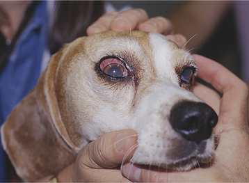
Figure 9-9 Beagle with primary open-angle glaucoma. “Red eye,” corneal edema, and dilated pupil are present.
Only gold members can continue reading. Log In or Register to continue
Stay updated, free articles. Join our Telegram channel

Full access? Get Clinical Tree


