Neurohistology, physiology and supporting structures

Chapter 3 Neurohistology, physiology and supporting structures


Neurohistology


Cells of the nervous system


The principal functional unit of the nervous system is the neuron. This is a highly specialised and polarised cell that receives, integrates and transmits information.


In the human brain it is estimated that there are 100 billion neurons. In contrast, the well-studied nematode, Caenorhabditis elegans, manages to go through life with a grand total of 302 neurons. There are five to ten times more neuroglia supporting the neurons. Neuroglia do not transmit action potentials but are vital for the normal function of the nervous system. Current research is identifying that they have significant roles in disease states.


Neurons


Neurons are the core components of the vertebrate brain, spinal cord and nerves. Neurons are excitable cells that receive and integrate incoming information from sensory receptors and other neurons, and transmit information to other neurons or effector organs. A typical neuron consists of a cell body or soma. It has a series of branching processes called dendrites that receive information from surrounding excitable cells or receptors. The output of the neuron is through a single process, the axon, although this may then split into a number of collateral branches.


The soma (plural, somata) contains a relatively large, round nucleus with a prominent nucleolus (Fig. 3.1A). The size of the cell body ranges from 5 µm to more than 100 µm, for interneurons and motor neurons innervating striated muscle, respectively. The soma contains a cytoskeleton made up of neurofilaments and neurotubules. The cytoskeleton extends into the dendrites and axon, setting the diameter of, and providing internal support for, these slender processes.


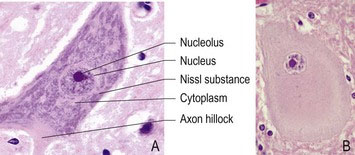
The soma contains most of the synthetic machinery of the cell, such as the Golgi apparatus and endoplasmic reticulum, while mitochondria are located in both the soma and axons. Nissl substance is granular material that stains with basophilic dyes; it is composed of rough endoplasmic reticulum. Nissl substance is the site of protein synthesis and its prominence in neurons indicates that they are highly metabolically active. In neurons that are damaged, especially after axonal injury, the Nissl granules are dispersed; this reaction is called chromatolysis (Fig. 3.1B).
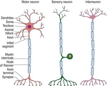

Most neurons lack centrioles, organelles involved in the organization of the cytoskeleton and the movement of chromosomes during mitosis. Consequently, most neurons cannot divide. One exception is the neurons of the olfactory bulb. These cells can divide and replace cells destroyed by pathogens contacting the olfactory mucosa. Olfactory neurons could provide stem cells for repairing injured CNS tissue; this is an area of active research. Neural stem cells exist in some areas of the brain, too, such as the subventricular zone of the forebrain. They may be the source of new neurons after birth.


Dendrites (déndron – Gk = tree) are the branched projections of a neuron that receive input from many other neural cells and conduct it to the soma. For example, the average mouse neuron receives input from 500 other neurons and has 8000 synapses. The input may be excitatory or inhibitory. The dendrites integrate the incoming information, and determine whether action potentials will be produced by the neuron. The dendrites of some neurons are covered with small membranous protrusion called dendritic spines. Each dendritic spine can, on occasions, synapse with multiple axons; thus one dendrite could communicate with hundreds of axons. Abnormal spines have been shown in the brains of humans with cognitive impairments. The dendritic branching pattern of a neuron can change and may increase or decrease. An enriched or stimulatory environment, such as when the animal is learning, is associated with the growth of dendrites.


Axons are cytoplasmic processes that can propagate electrical impulses. Axons may be only a few micrometres, as in interneurons, whilst axons in blue whales, they may be over 10 metres long. Neurites is a term that encompasses both axons and dendrites. Efferent neurites (axons) may be differentiated from afferent neurites (dendrites) in two ways: (a) efferent neurites usually maintain a constant radius, while afferent neurites often taper; (b) efferent neurites can be much longer than afferent neurites. This may not be the case for sensory neurons. For example a sensory neuron from a distal limb muscle, has a long afferent neurite, a cell body in the spinal ganglion and an efferent neurite that synapses in the adjacent spinal cord dorsal horn.


Owing to their length, axons usually contain the majority of the cell cytoplasm, and organelles such as neurofibrils, neurotubules, small vesicles, lysosomes, mitochondria and various enzymes. However, the majority of axonal proteins are synthesised in the soma, which may be located some distance from the distal portion of an axon. The axonal transport system is essential to carry material between the soma and the axonal tip. Microtubules (made of tubulin) run along the length of the axon and provide the main cytoskeletal tracks for axonal transport. The motor proteins, kinesin and dynein, move cellular cargoes in the anterograde (towards the axon tip) and retrograde (towards the cell body) directions, respectively. Motor proteins bind and transport several different cargoes including organelles such as mitochondria, cytoskeletal elements and vesicles containing neurotransmitters. Fast axonal transport is used to move organelles, structural proteins and neurotransmitters from their sites of synthesis in the cell body to the distant reaches of the axon at rates of hundreds of centimetres per day. Slow axonal transport moves components of the cytoskeleton at speeds of up to 2.5 mm/day. A retrograde transport system runs in the opposite direction, moving organelles from the distal axon back to the cell body at rates of up to 200 mm per day.


There are many types of neurons and they differ in the size of the soma, the length of the axon and the dendritic arborisation. Neurons are the most diverse kind of cell in the body with hundreds of different types, each with specific, message-carrying abilities. Neuronal types can be divided into three main groups:


Neuroglia


Neuroglia (glia – Gk = glue), are non-neuronal cells that provide support and nutrition, maintain homeostasis, form myelin, and participate in signal transmission in the nervous system. A number of types of neuroglia are recognised, principally astrocytes, oligodendrocytes, Schwann cells (collectively called macroglia) and microglia.


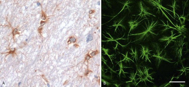
Astrocytes (Astro – Gk = star shaped) are a heterogeneous population of cells. Morphologically distinct examples of astrocytes include the protoplasmic astrocytes of the grey matter, fibrous astrocytes of the white matter and radial astrocytes of the retina and cerebellum. Most astrocytes express glial fibrillary acidic protein (GFAP) that is used as a cell-specific marker, histologically (Fig. 3.3). Astrocytes perform many functions, including contributing to the blood–brain barrier, regulation of blood flow, provision of nutrients to the nervous tissue, insulating synapses and maintenance of extracellular ion balance. Astrocytes have important roles in the repair and scarring process of the neuraxis following traumatic injuries or inflammatory disease. Bidirectional communication between astrocytes and neurons occurs, and their role in disease processes is increasingly being recognised.


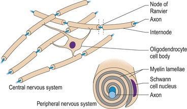
Oligodendrocytes (oligo – Gk = few branches) produce the myelin sheaths that surround many axons in the CNS. Schwann cells myelinate the axons in the PNS (Fig. 3.4). A single oligodendrocyte can myelinate one large diameter axon or up to 100 small-diameter axons, whereas Schwann cells only myelinate single axons. Myelin is essential for rapid, targeted conduction of nerve impulses. The myelin sheath is formed by outgrowth of cell processes from the myelinating cell, spiralling around the axon to form the numerous layers of membranes. Adjacent segments of myelin are called internodes. Internodes are separated by 1-µm gaps called the node of Ranvier. Ion channels clustered in the axonal membrane at the node are used to propagate action potentials along the axon. The myelin sheath is lipid-rich, providing effective insulation and blocking the exchange of ions across the axonal membrane in the internodal regions. Thus the action potential skips over the internodal areas as it jumps between nodes. This is saltatory conduction (saltus – L = to leap or bound). The myelin sheath also physically protects the axon. Its loss renders the axon vulnerable to chemical and mechanical damage.


Microglia are the resident macrophages of the CNS, and thus act as the first and main form of active immunity. Microglia comprise 20% of the total glial cell population within the brain and are constantly surveying the neuraxis for damaged neurons, plaques, and infectious agents. The brain and spinal cord are considered immune-privileged organs in that they are separated from the rest of the body by the blood–brain barrier (BBB) (see Fig. 3.19). This barrier prevents many pathogens from reaching the nervous tissue, but it also blocks most antibodies from accessing the neuraxis, due to their large size. Hence, microglia must be able to recognize foreign bodies, phagocytose them and act as antigen-presenting cells to activate other immune cells.


Aug 26, 2016 | Posted by in INTERNAL MEDICINE | Comments Off on Neurohistology, physiology and supporting structures

Full access? Get Clinical Tree

Get Clinical Tree app for offline access