Chapter 2 Neuroembryology
Key points
 During early embryonic development, the neuroectoderm forms the neural tube and neural crest; these structures become the central nervous system (CNS) and cells of the peripheral nervous system (PNS), respectively.
 The neural tube forms the spinal cord and brain, and the neural canal forms the inner ventricular system. The brain divides into prosencephalon, mesencephalon and rhombencephalon.
 Dorsal outgrowths of the neural tube give rise to the cerebrum and cerebellum. The degree of cerebellar development at birth determines the mobility of a neonatal animal.
 Germinal cells surrounding the neural canal give rise to neurons and glial cells; these cells migrate to form grey and white matter. The germinal layer becomes the ependyma in the postnatal animal.
 Neuronal differentiation is determined by extracellular morphogens. Sonic Hedgehog protein triggers motor neuron development in the ventral spinal cord, while Bone Morphogenetic Protein induces sensory neuron differentiation in the dorsal spinal cord. Different homeobox proteins induce the longitudinal differentiation of the spinal cord.
 Neuronal precursors migrate in response to molecular signals. Axonal growth is directed by chemoattractants or chemorepellants in the extracellular environment.
Development of the CNS
Formation of the neural tube
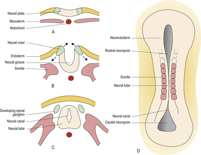
The notochord is derived from the mesoderm and establishes the cranial–caudal axis of the embryo. It induces the overlying ectoderm to become neurectoderm. The neurectoderm thickens to form the neural plate along the dorsal, longitudinal axis of the embryo. A longitudinal trough appears in the midline of the neural plate, forming the neural groove. The sides of the plate rise up and seal over the top of the groove to form the neural tube, which surrounds a fluid-filled, neural canal. Closure of the neural tube is called primary neurulation; it occurs at about 20 days in the canine fetus (canine gestation is approximately 60 days). Closure starts in the cervical region and extends rostrally and caudally from that point. The ends of the tube may remain open for a period as the rostral and caudal neuropores. Just prior to neural tube formation, neural crest cells develop, located dorsolaterally at the junction between the neurectoderm and the ectoderm. The neural crest cells separate from the neurectoderm and the ectoderm as the tube develops. Columns of neural crest cells form along the dorsolateral aspects of the neural tube, while the ectoderm fuses dorsal to the neural tube creating the overlying skin. The neural tube extends the length of the embryo including into the head region and forms the basis of the spinal cord and the brain. The neural canal will develop into the inner ventricular system (see Chapter 3). Inside the head, the neural tube develops into three regions, the prosencephalon (forebrain = telencephalon and diencephalon), the mesencephalon (midbrain) and the rhombencephalon (hindbrain = metencephalon and myelencephalon). The sulcus limitans is a groove that forms in the lateral walls of the neural canal. It demarcates the tube into dorsal/alar and ventral/basal plates that are associated with sensory and motor functions, respectively (Fig. 2.1).
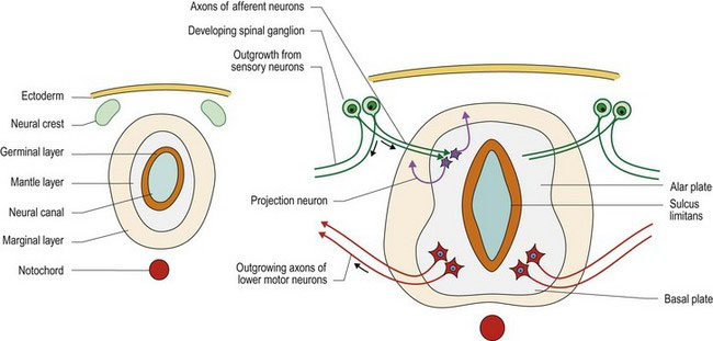
During differentiation of the neural tube, three cell layers develop from the original pseudostratified, single cell layer. Innermost, sited around the neural canal, is the germinal layer, in which cellular proliferation occurs giving rise to both neurons and the supporting glial cells, oligodendrocytes and astrocytes. The post-mitotic cells migrate so that the neurons are located in the middle or mantle layer, while their axons extend into the marginal layer (Fig. 2.2). Astrocytes are found in both the mantle and marginal layer, while oligodendrocytes settle in the marginal layer to myelinate the axons. After development has finished the germinal layer becomes the ependymal layer surrounding the ventricular system of the brain and the spinal cord. The mantle layer becomes the grey matter and the marginal layer becomes the white matter. The grey and white matter form continuous columns; these extend the length of the spinal cord and into the brainstem. Overall, the brainstem develops from the neural tube in a similar way to the spinal cord (see Figs. 1.7, 10.2). However, the columns of grey and white matter become fragmented with the grey matter forming nuclei. Additionally, in the rhombencephalon, the dorsal aspect of the tube opens back up forming a groove-like structure, but with a thin cover called the medullary velum (velum – L = awning). The neural canal is expanded to become the fourth ventricle. The tube reforms in the midbrain, with a small-diameter neural canal, the mesencephalic aqueduct (Fig. 1.7).
Stay updated, free articles. Join our Telegram channel

Full access? Get Clinical Tree


