Chapter 9 Nervous disorders
Introduction
The diseases and disorders covered in this chapter have predominantly nervous signs. Consequently, a wide etiological range is covered including nutritional conditions (e.g., cerebrocortical necrosis), metabolic disorders (e.g., hypomagnesemia), bacterial and viral infections (e.g., listeriosis and rabies), parasites (e.g., Coenurus cerebralis), physical and traumatic incidents (lightning strike and electrocution), and miscellaneous conditions of uncertain etiology (e.g., bovine spongiform encephalopathy). However, other diseases with significant clinical nervous signs may be featured elsewhere and include tetanus (12.66–12.68), botulism (12.69), and lead poisoning (13.29).
Cerebrocortical necrosis (polioencephalomalacia)
Etiology
cerebrocortical necrosis (CCN) is an induced thiamine deficiency caused by products of abnormal ruminal fermentation (thiaminases). Concentrate feeding leading to excessive growth of specific thiaminase 1 enzyme-secreting rumen bacteria is the most likely cause. The syndrome can also be induced by feeding high levels of certain ammonium sulfate mixtures used as dietary urinary acidifiers for the prevention of urolithiasis (10.5–10.10).
Clinical features
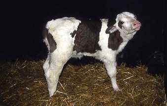
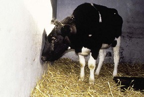
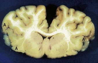
CCN is seen most commonly in calves that are 2–6 months old, often following a dietary change when on high-concentrate rations. The Simmental crossbred calf in 9.1 has a characteristic “star-gazing” stance. Signs can be very variable depending on whether the onset is acute or subacute. Other signs include depression, ataxia, head-pressing (9.2), and cortical blindness. Autopsy lesions (9.3) are normally symmetrical and occur in the frontal, occipital, and parietal lobes. Congestion and yellow degeneration of the cortical gray matter (A) is seen, typically at the junction of the white and gray matter, particularly on the left and right extremities. Affected brains will fluoresce blue-green under ultraviolet light.
Metabolic diseases
Hypomagnesemia (“grass staggers”, “grass tetany”)
Clinical features
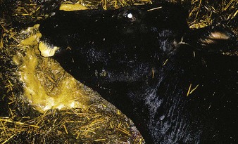
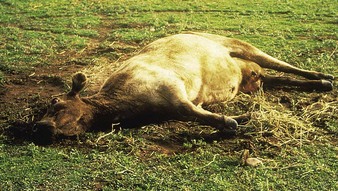
the Friesian cow in 9.4 fell and developed extensor spasm when being brought in for milking. Note the “staring” eye, dilated pupil, frothing at the mouth, and sweaty coat. In 9.5 the crossbred cow from Queensland, Australia, shows similar eye changes. The head and the hind legs are in extensor spasm. Violent paddling movements of the forelegs and head have resulted in loss of foliage, exposing the bare earth. Less severely affected cows may walk stiffly, are hypersensitive to touch and sound, and urinate often. Precipitated by stress and seen especially in temperate climates, the condition is induced by grazing magnesium-deficient or high-potassium pastures, and other pastures where magnesium uptake is poor. Concurrent hypocalcemia may be an exacerbating factor.
Differential diagnosis
hypocalcemia (9.6, 9.7), BSE (9.36–9.38) encephalitis, listeriosis (9.11, 9.12), ketosis (9.8).
Hypocalcemia (“milk fever”, postparturient paresis)
Clinical features
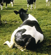
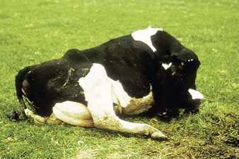
hypocalcemia (9.6) occurs typically in older cows immediately pre- or postcalving. Early signs include hypersensitivity and increased excitability. Later, affected animals are unable to rise owing to lack of muscle power and poor nerve function. Note also the protruding anal sphincter (due to accumulation of feces in the rectum and increased intra-abdominal pressure), slight ruminal bloat (ruminal atony), and the typical “S-bend” in the neck (9.6). This is thought to be a self-righting response, as the animal attempts to avoid full lateral recumbency. Some affected cows lie with their head resting on their flank (9.7).
Differential diagnosis
toxic mastitis or metritis, botulism (12.69), periparturient hemorrhage, severe hindlimb trauma (7.90), bilateral obturator paralysis (7.91).
Nervous acetonemia (ketosis, “slow fever”)
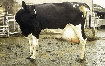
Nervous acetonemia is an intoxication by circulating ketone bodies and is associated with an energy deficit in early lactation. Typical clinical signs are anorexia and lethargy (hence “slow fever”), depressed yields, and constipation, although some cases develop nervous signs such as compulsive licking, salivation, biting flanks (as seen in this 5-year-old Holstein in 9.8), or even maniacal behavior. This cow was difficult to get in the parlor for milking. Six hours later her frantic licking of udder and forelegs resulted in bleeding. The cow rapidly responded to dextrose and corticosteroids.
Fatty liver syndrome (fat cow syndrome)
Clinical features
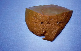
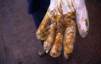
many cows show no specific clinical signs. More advanced cases develop anorexia and “star-gazing”, progressing to toxemia and terminal recumbency. Massive fatty infiltration of the friable liver is seen in 9.9. The plastic glove in 9.10 demonstrates the severe yellow scour typical of toxemia, which proved fatal in this dairy cow.
