Chapter 3 Integumentary disorders
Introduction
Visual appraisal of the skin is easily carried out and a wide range of disorders is recognized. Anaphylactic reactions can produce urticaria. Photosensitization may result from a range of intoxications including St. John’s Wort, Lantana, and facial eczema (see also Chapter 13, 13.13, 13.22–13.24). Parasitic (lice and mange), fungal (ringworm), bacterial infections (skin tuberculosis), and fly infestations (myiasis and warbles) all produce skin changes which are discussed in this chapter. The final section deals with physical conditions such as hematomas, abscesses, frostbite, other traumatic incidents, and neoplasia.
Many skin changes which are secondary to other diseases are described in the relevant chapters, for example, gangrene secondary to mastitis (see 11.8) or ergot poisoning (see 7.159), or subcutaneous swellings associated with urolithiasis (see 10.7) or umbilical (navel) conditions (2.9).
Cutaneous urticaria (urticaria, angioedema, “blaine”)
Clinical features
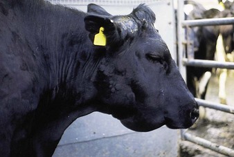
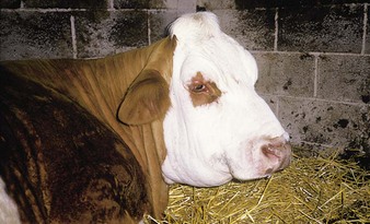
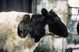
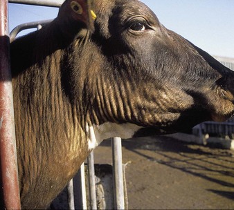
urticaria is sudden in onset. Cases are sporadic. The wheals do not itch. The Friesian cow (3.1) has raised plaques of edema (wheals) over the face and shoulders. The eyelids and muzzle are swollen. Although looking depressed, she was eating well and, like many such mild cases, recovered within 36 hours. The Simmental cow (3.2) was much more advanced, pyrexic, and in considerable pain. The head, grossly enlarged due to subcutaneous edema, was often rested on the ground. The skin of the muzzle was hyperemic. Localized areas sloughed a few weeks later. Some cases are allegedly due to snake bites or bee stings, but remain unproven.
Photosensitization (photosensitive dermatitis)
Clinical features
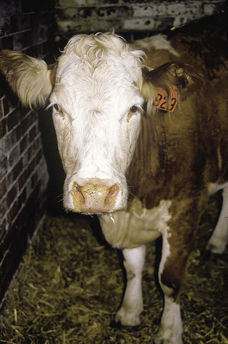
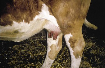
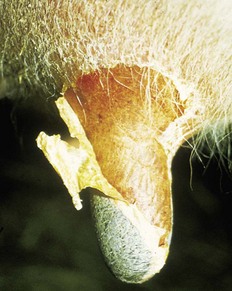
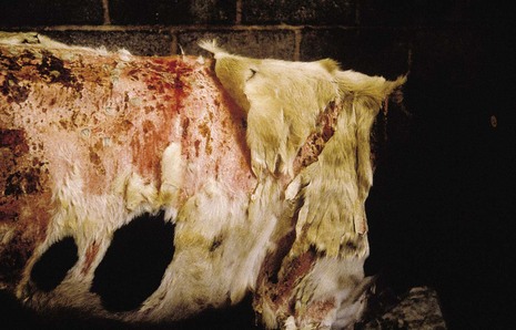
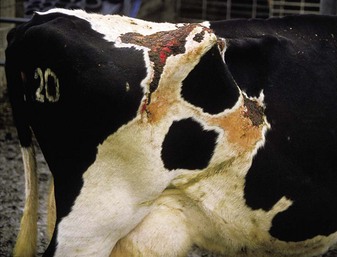
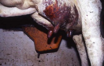
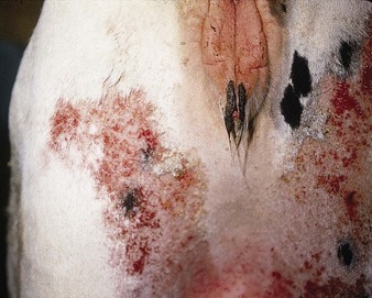
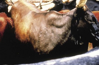
in the early stages, affected animals show marked discomfort and pyrexia, with erythema and encrustation around the margins of the nostrils (3.3) and erythema of the teats (3.4), as in this Simmental cow. The teats are very painful and may later blister and slough (3.5), making milking almost impossible. The thickness of the skin slough, in this case only moderate, depends on the degree of initial damage. The primary febrile phase, edema, and thickening of the white skin had passed unnoticed in the heifer in 3.6, and she was presented with sloughing of dry, hard areas of white skin and with a new epidermis forming beneath. Seven weeks later (3.7) the new skin was well developed. Hair regrowth was possible owing to the preservation of hair follicles deeper in the epidermis. Areas of granulation tissue may retard healing (3.8), especially over bony prominences such as the pelvis. A dry dermatitis persisted in this cow for a further 2 years. The condition also occurs in Lantana poisoning (13.13) and facial eczema (13.22–13.24). Serum biochemistry or hepatic biopsy may aid confirmation of liver damage. A more advanced case, involving both udder and teats, is shown in (3.9). The associated pain plus thickening of the teat wall (which prevented the teats filling with milk) made this cow impossible to milk.
Differential diagnosis
BEPP (1.35), foot-and-mouth disease (12.2–12.8), bluetongue (12.17), bovine herpes mammillitis (11.18), vesicular stomatitis (11.26, 11.27). The clinical presentation of early cases, where skin edema may be difficult to detect, strongly resembles colic.
Brown coat color
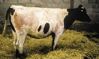
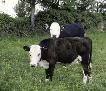
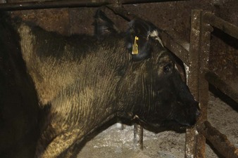
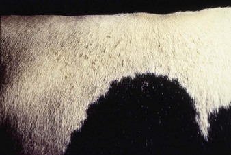
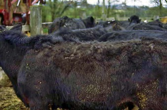
Copper deficiency affects several systems (see 7.167–7.172), but classically causes loss of coat color. However, copper deficiency is not the only cause of brown coat color. Animals turned out to spring grazing may retain their winter coat (3.10), particularly younger animals in poorer body condition and first-lactation heifers. Although grazing the same pasture, the older animal at the rear is not affected.
Mange
Sarcoptic mange (scabies)
Caused by Sarcoptes scabiei var. bovis, lesions are typically seen over the head, neck and hindquarters (3.11). Note the hair loss and severe thickening of the skin in 3.12. The white areas show secondary damage due to rubbing. In severe cases there may be almost total hair loss. The close-up view (3.13) shows the dry, scaly appearance of the thickened skin.
Chorioptic mange
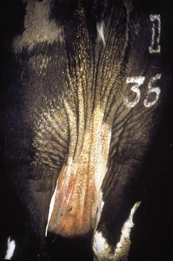
Chorioptic mange is the most common type of mange in cattle. The fold of skin beside the tail is the characteristic site for infestation by Chorioptes bovis (3.14). Lesions comprise a thick encrustation overlying an area of moist serous exudate. They are intensely irritant. In untreated cases the dry encrustation spreads down the perineum and over the posterior udder. In advanced cases (3.15) red, pustular lesions may be seen. Less commonly infection is seen in the neck and shoulder region (3.16).

Psoroptic mange
Note the skin thickening and hair loss in the cow (3.17), extending from the vulva to the udder. The condition may start at the withers and spread over the whole body. Pruritus is often marked. Psoroptic mange (Psoroptes ovis) is notifiable in North America, where eradication programs have been in progress for many years. In recent years psoroptic mange has also become more common in South Wales and elsewhere in Europe, especially Belgium.
Demodectic mange (follicular mange)
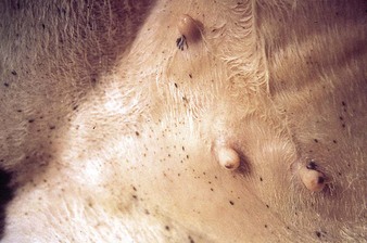
Small papules are seen on the white skin of this cow (3.18), from which a thick, white, waxy material containing large numbers of mites can be expressed. Another case (3.19) shows papules over the skin of the shoulder. Some nodules become secondarily infected with Staphylococci. The condition is generally mild, with a spontaneous recovery. Extensive hair loss is rare. Similar lesions are seen with Dermatophilus (3.38, 3.42).
Lice (pediculosis)
Clinical features
sucking lice (e.g., Haematopinus eurysternus and Linognathus vituli and, in North America, Solenopotes capillatus) are slower moving than biting lice (Damalinia [Bovicola] bovis). In addition to pruritus (the only lesion due to biting lice), sucking lice may produce severe anemia, loss of condition, and even death. Pediculosis is most prevalent in the winter months. Parting the hair along the back may reveal small, brown, motile lice just visible to the naked eye. They are more easily seen on white skin and hair, or on the hairless skin of the groin (3.20). Note the variation in their size.
Clinically, infestations are manifested by rubbing, biting, scratching, and thickening of the skin. In 3.21 the calf’s tongue is protruding and its head is held on one side, a stance typical of pruritus. In early cases the hair develops vertical lines on the neck. Small, hairless areas with white scurf may arise from biting. In more advanced cases the skin is thickened round the face and the vertical hair lines on the neck are thrown into thick folds, as in this adult Ayrshire cow (3.22). A further characteristic sign is the presence of clumps of hair (3.23) where affected cattle have been biting their pruritic skin, as opposed to licking and grooming it.
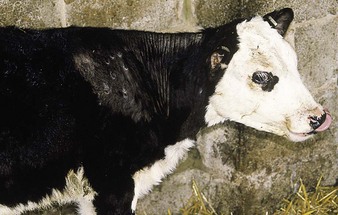
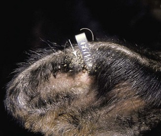
Younger calves especially may be stunted and anemic, and become much more susceptible to other diseases such as pneumonia, especially with sucking lice infestations. Ringworm often occurs in association with lice: early lesions are seen on the shoulder in 3.21. Pale beige-colored oval lice eggs (“nits”), glued onto the hair shafts, can often be seen with the naked eye, particularly on the ear (3.24 near the tag) and over the shoulders. In older animals the coat may become matted with lice eggs.
Ringworm (dermatophytosis)
< div class='tao-gold-member'>
Stay updated, free articles. Join our Telegram channel

Full access? Get Clinical Tree


