Chapter 2 Neonatal disorders
Introduction
This chapter covers disorders of the calf from birth until postweaning. The first section deals with navel ill, umbilical hernia, and general conditions of the navel. Later sections cover different forms of diarrhea and alopecia, with a miscellaneous group including calf diphtheria and joint ill. According to the presenting signs, other diseases of calfhood are considered in the relevant chapters; for example, lice, ringworm, and skin diseases are to be found in Chapter 3, respiratory problems in Chapter 5, and meningitis in Chapter 9.
Conditions of umbilicus (navel)
Umbilical eventration
Clinical features
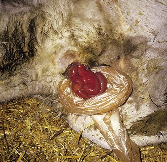
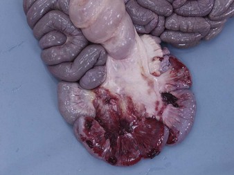
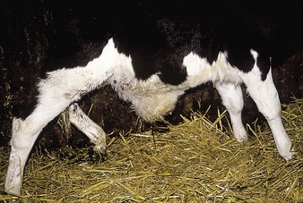
umbilical eventration is seen in a small proportion of calves immediately after birth. The prolapsed intestines (jejunum) may be fully exposed, as in the Friesian (2.1), or contained in a sac of peritoneum. Opening the sac in a Charolais calf revealed a congested intestine (2.2). Often the exposed intestine ruptures when the calf moves. The prognosis is then hopeless. In more advanced and exposed cases the intestinal loops turn a deep red/purple color due to ischemic necrosis (2.1).
Navel ill (omphalophlebitis)
Clinical features
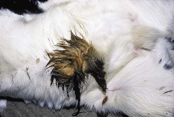
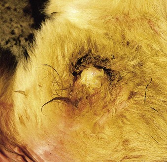
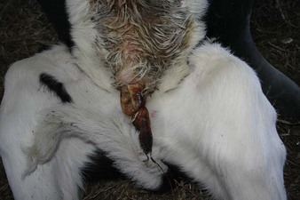
lacking skin or any other protective layer, the moist, fleshy navel cord is particularly prone to infection until it dries up, normally within 1 week of birth. In the first calf (2.3) (shown at 3 days old) the enlarged and still moist navel cord is seen entering an inflamed and swollen umbilical ring. Navel ill is uncommon at this age.
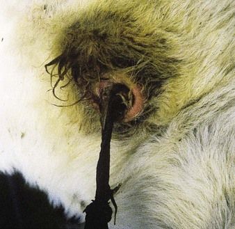
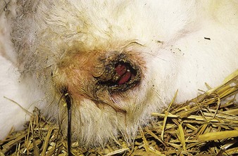
The more typical case is pyrexic, with a swollen, painful navel exuding a foul-smelling creamy-white pus (2.4). Culture usually reveals a mixed bacterial flora including Escherichia coli, Proteus, Staphylococcus, and Arcanobacterium pyogenes. This case persisted for several weeks.
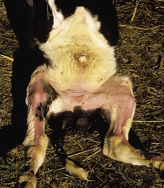
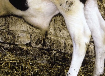
Alopecia on the medial aspects of the thighs (2.5) is due to a combination of urine scald and excessive cleansing of the navel by the owner. Some cases show no gross discharge, but the tip of the swollen navel will be moist and malodorous.
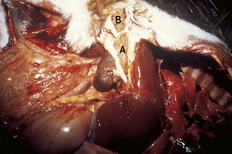
In other cases an intra-abdominal abscess may develop in the omphalic (umbilical) vein. In 2.6, A shows the intra-abdominal abscess in the grossly distended umbilical vein, adjacent to the navel, B. Spontaneous rupture of the abscess can lead to death from peritonitis, as in this calf. Occasional cases involve the urachus to produce a cystitis which can lead to stunted growth, sickness, and death several months after birth.
Septicemia can result in localization of infection in the joints (2.48, 2.49), meninges, endocardium, or end-arteries of limbs.
Umbilical granuloma
Definition
a tumor-like mass of granulation tissue due to a chronic inflammatory process at the navel.
Clinical features
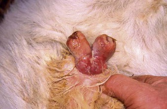
a small bifurcated mass of granulation tissue protrudes from the navel of this 2-week-old calf (2.7). Many cases consist of a single mass of tissue. It is only slightly painful and affected calves are generally not pyrexic, although there may be superficial infection present, as in this case. The condition will not resolve until the mass is removed by ligation at its base. If left untreated (2.8), it will persist into adult life.
Umbilical hernia
Clinical features
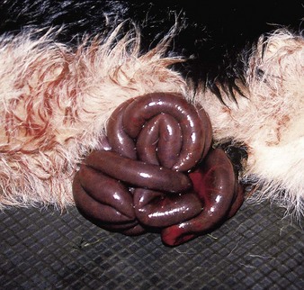
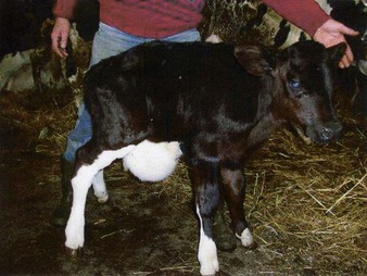
a large, soft, and fluctuating ventral abdominal swelling can be seen in this 3-month-old Friesian calf (2.9). The arched back in this calf clearly depicts discomfort. Many smaller hernias are not associated with pain. Rarely, in moderate-sized umbilical hernias, the jejunal loops become strangulated, and the calf is in severe pain, becomes toxemic, and dies within a day or two. Autopsy of one such case (2.10) shows the multiple loops with severe serosal hemorrhage. Although present from birth, some hernias are not noticed until the calf is at least 2–3 weeks old. A proportion of cases are inherited. Irreducible and strangulated hernias are uncommon.
Umbilical abscess
Clinical features
the swelling in this 4-month-old Friesian male (2.11) is cranial to the prepuce (compare urolithiasis (10.7), where it is caudal), and appeared spontaneously. The mass was initially hard, hot, and painful. Pyrexia led to systemic illness. Parenteral antibiotics resulted in a change to a more fluctuating swelling, which was successfully lanced and drained.
A hernia and an umbilical abscess can occur together. Occasionally, navel ill or navel abscess produces a localized peritonitis that erodes through the rumen wall to produce a rumenal fistula. 2.12 shows a 3-month-old Friesian male with a grossly enlarged navel sac, soiled anteriorly. Rumen contents leaked through the fistula, shown in close-up in 2.13.
Navel suckling
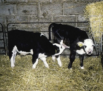
Navel suckling (2.14) is a common vice in group-housed, bucket-fed calves, especially if they are in poor condition and have intercurrent diseases. The calf being sucked has an enlarged navel, which could be infected. There is hair loss around the navel, indicating a chronic problem. The ears, tail, and scrotum can also be suckled.