Chapter 8 Ocular disorders
Congenital disorders
Although by definition congenital abnormalities are present at birth, some may not be recognized until the calf is much older. Strabismus (squint) is a typical example. Congenital disorders may be genetic, and therefore inherited, or they may be caused by environmental factors. Some abnormalities have more than one cause. For example, congenital cataract may be inherited, or it may have been caused by maternal BVD infection during pregnancy. The cause of many abnormalities is unknown. Congenital disorders in organs other than the eye are described in Chapter 1.
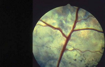
BVD/MD, discussed under “Alimentary disorders” (p. 54) can give rise to congenital or acquired ocular changes. Congenital BVD/MD can cause teratogen-induced retinal necrosis and degeneration, focal capsular cataract, as well as optic nerve gliosis, microphthalmos (see below) and optic neuritis. 8.1 shows retinal changes in a calf associated with the teratogenic effects of BVD/MD virus. There is marked attenuation of many retinal blood vessels, retinal hyper-reflectivity and a patchy yellow pigmentary disturbance.
Anophthalmos (anophthalmia); microphthalmos (microphthalmia)
Clinical features
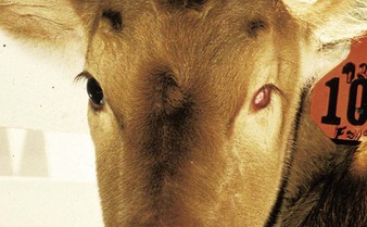
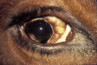
the two examples illustrate both abnormalities. The left eye of the Guernsey heifer in 8.2 has a small orbit and there is no evidence of the globe. Note that the entire orbit appears collapsed and smaller, compared with the normal right eye. This condition can be inherited. A Jersey cow with microphthalmos (8.3) and prolapse of orbital fat had possibly had an insult to the eye in calfhood, leading to this shrunken globe (phthisis bulbi).
Cataract
Clinical features
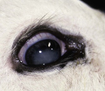
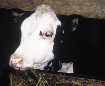
both eyes of the 4-day-old Hereford crossbred calf in 8.4 were affected and the animal was totally blind. In other animals, only one eye may be affected, or the cataract may not cause total loss of vision. Congenital cataract is not normally progressive. Cattle cope with blindness remarkably well and can be reared in confinement systems. They quickly learn to remain within the group, although handling can be difficult. Blind dairy cows will learn to follow the herd to and from pasture. Congenital cataract may be inherited, or may result from the teratogenic effects of maternal BVD infection during early/mid pregnancy. 8.5 shows a congenital nuclear cataract in a young Friesian calf.
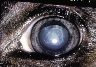
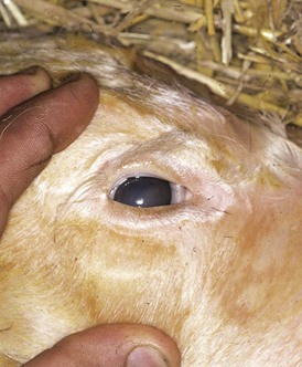
Note in acquired cataract the two large synechiae (adhesions of the iris to the cornea), and the opacity and wrinkling of the lens in the Guernsey cow in 8.6. Cataracts may be secondary to inflammatory processes within the eye, when they can be progressive. In contrast, congenital cataracts (8.4) are rarely progressive.
Coloboma
Definition
a coloboma is a congenital cleft caused by failure of the embryonic optic fissure to close.
Dermoid
Clinical features
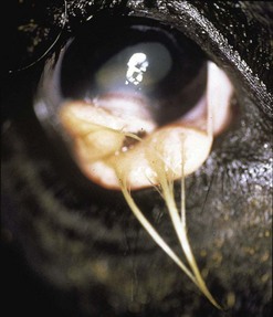
a typical dermoid is seen in a 4-month-old Friesian heifer (8.8). The tumor is attached to the conjunctiva of the lower lid and presents long hairs, which led to the presenting sign of unilateral epiphora.
Strabismus (“squint”)
Clinical features
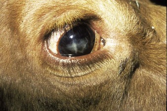
strabismus may be convergent (esotopia), when the visual axes of the eyes converge more than is required for normal vision, or divergent. It may involve one or both eyes. The globe is deviated from its proper axis due to excessive tension in opposing extraocular muscles. 8.9 shows convergent strabismus in the left eye of a Hereford cross heifer. Exophthalmos with strabismus may be inherited, although it is often unnoticed until 6–9 months old, and it is frequently progressive. Some animals may become so badly affected that total impairment of vision results.
Acquired disorders
Vitamin A deficiency
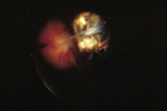
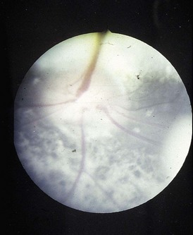
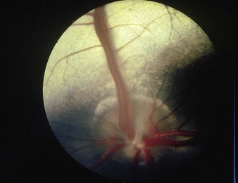
In young growing animals, vitamin A deficiency blindness is associated with stenosis of the optic foramen and consequent pressure on the optic nerve. The pupil becomes dilated and degenerative changes may be seen on the retina (8.11). For comparison, 8.12 shows a normal fundus. The optic disc is pale and enlarged, with indistinct margins (papilledema). White mottling of the nontapetal area suggests chorioretinal degeneration. The steer was blind. The diet had been barley straw, rolled barley and, occasionally, poor-quality hay.
< div class='tao-gold-member'>
Stay updated, free articles. Join our Telegram channel

Full access? Get Clinical Tree


