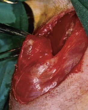
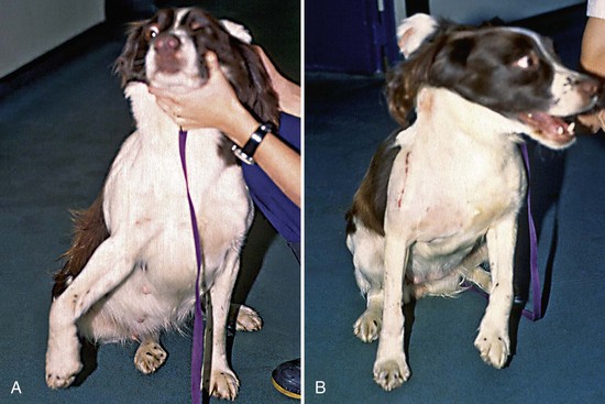
Chapter 70 Muscle and tendon disorders in animals present a unique challenge in veterinary practice. Until recently, muscle disorders could be diagnosed only by clinical examination. A survey of veterinary teaching hospitals revealed that less than 5% of dogs presenting for musculoskeletal disorders were classified with muscular disease or injury.35 This report directly contrasted with findings in human medicine, in which muscle problems represent the most common injury seen in general practice,36 and chronic painful muscle problems are a clinical problem without an easy solution.34 In the animal population, increased use of advanced imaging techniques, including ultrasonography, computed tomography (CT), and magnetic resonance imaging (MRI), is improving the veterinarian’s ability to diagnose muscle disease or injury. Muscle injuries can arise as a result of direct trauma or athletic activity, or as a complication of surgical procedures. Although injuries of muscles are common, they rarely cause a significant clinical problem because full recovery after injury is the usual outcome. Exceptions to this are specific injuries in athletic dogs (e.g., racing Greyhounds) and situations in which muscle contractures develop.25 Muscle injuries are commonly classified as contusions (blunt injuries), strains (indirect injuries), or lacerations (sharp injuries).28 Injuries to the musculotendinous unit usually result from an overstretching or strain injury, by far the most common type, or from direct trauma in association with other injuries such as fractures. In fractures, the energy released as the bone breaks is absorbed by the muscle, and additional injury occurs as a result of lacerations or tears from sharp bone fragments. Muscle can also be lacerated as the result of external trauma in the absence of fracture. Significant damage can be caused by iatrogenic injury sustained during surgical procedures. Muscle strains most commonly occur at the musculotendinous junction,29,56 although they can arise at the origin, at the insertion, or through the belly of the muscle itself. Strains may result after a powerful active contraction of the muscle occurs simultaneous with passive extension of the muscle unit.50 Muscles that cross two or more joints appear to be more prone to injury. Various degrees of damage, ranging from mild bruising to full rupture of the muscle, can occur; the degree of injury depends on the forces generated. Injuries are classified into the following three stages to aid prognosis and management.32 Stage I: myositis and bruising but architecture intact Stage II: myositis and some tearing of the fascial sheath Stage III: tearing of the fascial sheath, muscle fiber disruption, and hematoma formation Muscle healing occurs through a combination of two processes: direct regeneration of myofibrils and production of fibrous scar tissue. The functional result depends on a balance of these two important processes. This balance is determined by several factors, including the source of the myoblasts, an intact extracellular matrix, adequate vascularization, adequate innervation, and limited stress across the healing wound. Lack of any of these factors can lead to a predominance of scar tissue.44 Myofibrils regenerate rapidly, provided the sarcolemmal nuclei have survived, and can effect a quick and complete repair if the endomysium is intact. If the sheath is damaged, regenerating myofibril heads will form many buds that will attempt to cross the damaged area. Formation of excess fibrous tissue impedes this and impairs the effectiveness of the final result, possibly leading to recurrent injury.48,50 Immediately after injury, damage to the local blood vessels and possibly to myofibrils and the muscle sheath may be noted, depending on the severity of the injury. Hematoma formation is followed by an inflammatory response, with cellular infiltration and phagocytosis occurring 6 to 12 hours after injury. The healing process commences within 48 hours with invasion of capillaries and myoblast proliferation, followed by myofiber formation. Fibroblast proliferation and collagen scar formation are evident from days 4 to 6, filling the damaged area with a new collagen network by day 10. Tissue strength increases rapidly up to day 14, when the entire process slows down.24 The primary objective of treatment is to maximize the direct myofibril repair while minimizing scar formation. Early diagnosis and prompt intervention lead to optimal results. In situations in which healing occurs with excessive scar production, the efficiency of the muscle is compromised and the ability to produce tension can be reduced to 50% of original capability.27 In contusions, as well as in stage I and many stage II strains, good healing can be expected with direct growth of new muscle tissue.15 The aim of management is to minimize the early disruptive effects of inflammation, edema, and swelling. In the first 24 to 48 hours after injury, cold compresses and nonsteroidal antiinflammatory agents are indicated.10,49 Light compression bandages can be used if anatomically feasible.10 Early mobilization is essential for proper myofibril orientation and can be considered after 5 to 10 days, once the initial injury has settled. Too much mobility too soon can result in additional damage. However, muscle often remains strong enough to repair well and resist further serious damage with controlled mobilization even after significant disruption.57 In advanced stage II and in stage III injuries, early surgery should be instigated to eliminate any gaps and to appose the muscle to optimize healing without scar.24 Surgery should be planned once the initial inflammatory phase has receded; this typically occurs 2 to 3 days following injury. Until this point, the injury should be treated with cold compresses and nonsteroidal antiinflammatory medication. Evacuation of the hematoma and debridement of any necrotic material are followed by muscle apposition. Great care should be taken in handling the damaged muscle to prevent additional trauma. Monofilament long-term absorbable suture material is used to place deep horizontal mattress tension sutures while the ruptured muscle ends are held apposed. These should be placed in the fascial layers for better retention and may be augmented with buttons or stents to prevent the suture from tearing through the muscle tissue.9 Once sufficient tension sutures have been applied to hold the bulk of the muscle in place, additional appositional sutures of smaller gauge monofilament long-term absorbable suture material are applied through the fascial sheaths at the damaged muscle ends.10 Rupture of the Long Head of the Triceps Brachii Muscle Avulsion of the origin of the long head of the triceps brachii muscle is a recognized problem in racing Greyhounds. A depression is present caudal and distal to the scapula. Conservative management of this problem is common and can result in healing, but the dog may not reach the same performance standards as it attained before injury. Repair can be performed by reattaching the muscle belly to the scapula.9 Rupture of the Gracilis Muscle Rupture of the gracilis muscle22 is a common and devastating racing injury in Greyhounds. It has also been described in German Shepherd Dogs and Foxhounds.39 Rupture can occur at the musculotendinous junction, or the origin or insertion can avulse from its attachment (Figure 70-1).9 Clinically, a large hematoma is evident on the medial aspect of the thigh, accompanied by bruising. A depression may be evident if a complete or significant rupture has occurred. Surgical repair or reattachment is the preferred treatment method.22 The iliopsoas and pectineus muscles have been identified as the most common muscles presenting with strain injury in the hindlimbs of pet dogs.13,47 Affected dogs may present with acute or chronic lameness of one hindlimb. Diagnosis is based on clinical evidence of pain on direct palpation of the muscle. Extension and internal rotation of the hip result in a pain response, as does digital pressure applied over the area of attachment of the iliopsoas muscle on the lesser trochanter. One study47 of dogs with hindlimb muscle strain injury implicated the iliopsoas muscle as the main cause of pain in 32% of cases; 23% had pectineus muscle strain, and 14% had strain of an unspecified adductor muscle of the hindlimb. In 25% of the cases described, a combined iliopsoas and pectineus muscle strain was diagnosed. All dogs were treated conservatively using a combination of rest, short-term administration of a nonsteroidal antiinflammatory medication, a muscle relaxant, and physical therapy, with most achieving resolution or significant improvement at follow-up. Muscle contractures are occasionally encountered in clinical practice and can be extremely difficult to treat. Muscle contracture is defined as “the abnormal shortening of muscle tissue, rendering the muscle highly resistant to stretching.”8 Shortening usually occurs as part of replacement of the majority of muscle tissue by fibrous connective tissue.58 Muscles can consist of a great deal of fibrous tissue and still function normally. Clinical signs represent a functional problem rather than a pathologic description. Many causes have been implicated, including compartment syndrome, infection, trauma, repetitive strain injury, fracture, prolonged immobilization, and various primary muscle diseases. Those muscles most commonly affected are the infraspinatus, quadriceps femoris, gracilis, and semitendinosus muscles. Other muscles in which clinical problems have been produced by contracture include the sartorius,43 supraspinatus,18,60 teres minor,14 iliopsoas,2 and brachialis58 muscles. Contracture of the infraspinatus muscle (see Chapter 51) is most commonly diagnosed in dogs.16,19 A contracture usually occurs unilaterally, but bilateral contracture has been described.26 Contracture is most commonly identified in medium-sized working or athletic dogs. Transient forelimb lameness often appears 4 to 6 weeks before onset of the physical signs of contracture. Clinically, the condition is characterized by weight-bearing lameness or a gait abnormality. When observed carefully, this lameness involves circumduction of the affected forelimb as it is advanced; this can be accompanied by a carpal flip. The dog is reported as being pain free and usually is still keen to work or exercise despite the lameness. This lack of obvious discomfort can result in delayed presentation for veterinary attention. When standing, the dog has its shoulder abducted, elbow adducted, and lower limb held abducted and externally rotated (Figure 70-2, A).56 The cause of the fibrotic contracture is unknown. Trauma is usually suspected, although an interesting theory suggests that the contracture may be the result of a localized compartment syndrome, leading to ischemic and neurologic damage.54 Surgical exposure and tendinectomy of the tendon of insertion of the infraspinatus muscle, along with release of surrounding fibrous adhesions, is the treatment of choice. This will result in an immediate return to normal gait, which is maintained over the long term (Figure 70-2, B).
Muscle and Tendon Disorders
Disorders of Muscle
Muscle Injury and Healing
General Principles of Treatment
Surgical Management
Muscle Injuries In the Forelimb
Muscle Injuries In the Hindlimb
Iliopsoas and Pectineus Muscle Strain
Muscle Contracture and Fibrotic Myopathy
Infraspinatus Muscle
![]()
Stay updated, free articles. Join our Telegram channel

Full access? Get Clinical Tree


