7 Malassezia dermatitis
CASE HISTORY
• A diagnosis of atopic dermatitis had been made 1 year previously and was based on history, clinical signs, failure of response to an appropriate diet trial and intradermal reactivity to environmental allergens (house dust mites, storage mites and mould allergens).
• Response to allergen-specific immunotherapy was poor but remission of pruritus had been successfully achieved with ciclosporin (5 mg/kg on alternate days).
• The flare-up had a duration of about 3 weeks, which did not coincide with the animal’s regular visits to a grooming parlour.
CLINICAL SIGNS
Malassezia dermatitis is less common in cats. It has been associated with facial dermatitis and with otitis externa secondary to allergic skin disease. Generalized lesions in cats have been associated with immunedysregulation in conditions such as the paraneoplastic syndromes.
In this case, the clinical signs included:
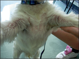
• Hyperpigmentation, lichenification, crusting and erythema on the axilla and the anterior aspects of the elbows (Fig. 7.1).
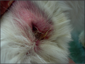
• Convex aspects of both ear pinnae and vertical ear canals were erythematous and oedematous (Fig. 7.2).


