Cheyletiellosis in a rabbit

16 Cheyletiellosis in a rabbit


INTRODUCTION


Ectoparasite infestation is a common problem in rabbit veterinary practice, with the most common parasites, Cheyletiella sp. and Leporacarus sp., both being referred to as rabbit fur mites. With cheyletiellosis, affected individuals usually have Cheyletiella parasitovorax, although C. takahasii, C. ochotonae and C. johnsoni have also been reported.


Cheyletiella is an obligate non-burrowing mite that feeds on the keratin layer of the epidermis, creating pseudo-tunnels through the scale and debris on the skin surface. The mite may also pierce the skin with its needle-like mouth parts to feed on tissue fluids. Transmission is by direct contact. The entire life cycle of Cheyletiella takes place on the host and is completed in 14–35 days. Despite preferring to remain on the host, adult females can survive off the rabbit host for approximately 10 days.


Clinical signs of cheyletiellosis are variable. Many rabbits will harbour the mite with no overt signs of skin disease but, where present, clinical signs are usually confined to mild pruritus with large soft white flakes of scale, with occasional alopecia, in the interscapular region or other dorsal body surfaces. Mites are just about visible to the naked eye and in moderate to severe infestations rabbits may appear to have ‘walking dandruff’. The disease is more common in young or immunosuppressed individuals and in animals suffering from an underlying condition that prevents grooming.


Diagnosis is via microscopic examination of acetate tape strips, or coat brushings, and treatment is fairly straightforward unless there is a more serious underlying disease.


Another common ectoparasite of rabbits is the other rabbit fur mite, Leporacarus gibbus (formerly named Listrophorus gibbus). It is also found in the fur of rabbits and infestations are often asymptomatic. If clinical signs are present they might include alopecia, seborrhoea and scaling. As with cheyletiellosis, an infestation severe enough to cause clinical signs is usually associated with an underlying disease process. The diagnosis and treatment of Leporacarus sp. is the same as for Cheyletiella sp.


CASE HISTORY


Complete and thorough history taking is very important. Much of the information obtained may not initially seem related to skin disease or the clinical presentation. However, over 75% of problems seen in rabbits and other small mammal pets are due to improper housing, diet, environment and general poor husbandry. This in turn will lead to general ill thrift, immunocompromise and increased susceptibility to ectoparasite infestation. The presenting signs may simply be the consequence of a more serious underlying disease such as dental problems or osteoarthritis (as discussed in the ‘Anatomy and physiology refresher’ section).


The relevant case history in this case was:


CLINICAL EXAMINATION


Prior to focusing on the dermatological problems, a full physical examination should be carried out to evaluate for any concurrent disease or additional abnormalities. In this case, the general physical examination was unremarkable, with only dermatological abnormalities noted. Examination also included otoscopic dental examination to evaluate the teeth, which was unremarkable, although not entirely reliable (see ‘Clinical tips’ section). Oral pain associated with dental problems is very common in pet rabbits. This pain will reduce grooming and can be an underlying cause for a dermatological problem.


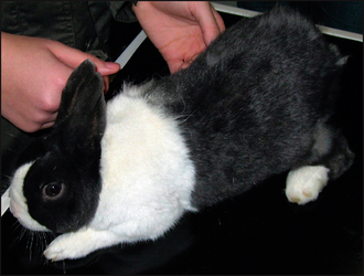
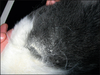
The dermatological examination in this case confirmed an area of marked scaling dorsally between the scapulae, although there was also mild scaling extending caudally along the dorsal midline of the back (Figs 16.1 and 16.2). The skin in these areas was mildly erythematous and there was thinning of the coat in the worst affected area.


The clinical signs of cheyletiellosis can be variable and are generally not severe. Many rabbits can harbour the mite asymptomatically, with no detectable signs of skin disease. In individuals that develop lesions, they are initially often subtle and may lead to a delay in seeking veterinary advice.


When these patients are eventually presented to a veterinary surgeon, the clinical signs are usually confined to large soft white flakes of scale in the inter-scapular region. There may occasionally be mild pruritus, but this is not a consistent feature. The mites can often just about be seen with the naked eye as they move about in the loose scale, giving rise to the descriptive term of ‘walking dandruff’. Depending on the duration and severity of the infestation, crusting may also be present, with alopecia and moderate to marked pruritus.


Ectoparasite infestation is more common in young or immunosuppressed rabbits, or those suffering from an underlying condition that reduces or prevents grooming (see ‘Anatomy and physiology refresher’ section).


CASE WORK-UP


Ectoparasites are a very common cause of dorsal scurf in rabbits, so microscopic examination of the scurf is indicated. As previously mentioned, it is possible to visualize Cheyletiella sp. with the naked eye, but this should not be relied upon for a definitive diagnosis.


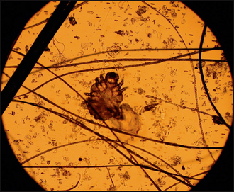
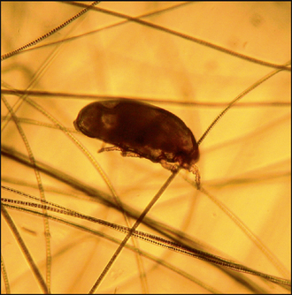
Acetate tape strips: The rabbit was gently restrained and two acetate tape strips were taken from the area of dorsal scale. These were mounted on a microscope slide and examined under the low-power objective (×40 magnification). Mites are found in greatest numbers over the scapulae, but may also be encountered on the back of the head, the neck and rarely on the caudal abdomen. Cheyletiella mites are typically saddle shaped with hook-shaped mouth parts and are easily seen using the described technique (Figs 16.3 and 16.4). Specific identification of the mite is not required in practice, and the vast majority found on rabbits will be C. parasitovorax.


It is useful to repeat the acetate tape strips in different affected areas to increase the likelihood of finding parasites. The sample should also be evaluated for other parasites such as Leporacarus sp., lice and fleas.


Cheyletiella mites were found in this case and further examination of the tape strips did not reveal any other ectoparasites. A negative result on acetate tape strips may have prompted further diagnostic tests.


Coat brushings: Examinations of coat brushings, or collections of scale, mounted on a slide might be equally useful in diagnosing cheyletiellosis. The scale should be collected and placed on a slide with liquid paraffin, then covered with a coverslip. The slide should be examined under low power, as with the acetate tape strips.


Skin scrapes: Occasionally, superficial skin scrapes may be required to demonstrate Cheyletiella sp. Skin scrape samples would also need to be obtained to evaluate for burrowing mites such as Sarcoptes sp. or follicular mites such as Demodex sp., since these are less likely to be found in the scale.


Dermatophyte culture: Dermatophyte culture may be carried out if multiple evaluations for ectoparasites were negative. Trichophyton mentagrophytes is common in outdoor rabbits, whereas Microsporum canis and M. gypseum are more common in pet and house rabbits. The presenting signs and the demonstration of Cheyletiella mites in the scurf made dermatophytosis unlikely in this case.


Blood sampling: Since cheyletiellosis may be associated with immunosuppression or an underlying disease, samples may be obtained for haematology and biochemistry (see ‘Clinical tips’ for help with blood sampling). This will provide the clinician with an overview of the patient’s health status. Serology for Encephalitozooan cuniculi would also be useful in cases that are complicated or refractory to treatment. Routine haematology and biochemistry were unremarkable in this case. Encephalitozooan cuniculi serology was negative.


Radiography: Since underlying disease is common, radiography of the thorax, abdomen and skull (for dental disease) would be indicated in complicated cases or those unresponsive to treatment.

Stay updated, free articles. Join our Telegram channel

Sep 3, 2016 | Posted by in SMALL ANIMAL | Comments Off on Cheyletiellosis in a rabbit

Full access? Get Clinical Tree

Get Clinical Tree app for offline access