Local and Regional Analgesic Techniques in Camelids
1. Because local anesthetic drugs provide complete pain relief from the nerves that are desensitized or “blocked,” many procedures can be performed under sedation and the sensory blockade alone, without the need for general anesthesia.
2. If general anesthesia is necessary, the sensory blockade provides analgesia during the procedure, allowing the patient to be maintained at a lighter plane of anesthesia and alleviating dose-dependent side effects of maintenance anesthetic drugs.
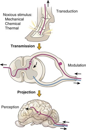
3. The likelihood of “windup” or central sensitization is greatly decreased because the portion of the pain pathway called transmission is blocked by local anesthetic drugs (Figure 48-1). Transmission involves the conductance of pain impulses from the peripheral nociceptors to the dorsal horn neurons in the spinal cord, which are primarily responsible for central sensitization. By blocking input to these neurons, central sensitization is less likely to occur.

Figure 48-1 Components of the pain pathway. When a painful stimulus is applied, pain receptors or “nociceptors” are activated in the periphery (“transduction”) and the painful impulse is moved up the peripheral nerves (“transmission”) to the dorsal horn of the spinal cord where dorsal horn neurons are activated by the release of excitatory neurotransmitters (“modulation”). The pain impulse is then carried up spinal tracts to the brain (“projection”) and, in the final phase of the pain pathway (“perception”), the patient becomes cognizant that pain has occurred. (From Anderson DE, Muir WW: Pain management in cattle, Vet Clin North Am: Food Anim Pract 21(3):623-635, 2005, with permission.)
The only criticism about local anesthetics is that the duration of action of these drugs is relatively short compared with the duration of pain from surgery, trauma, and so on. Thus, local anesthetics should be combined with other drugs, either at the site of locoregional blockade or systemically, for maximum analgesic efficacy.
Drugs Used for Locoregional Analgesia
Most of the locoregional blocks described in this chapter are achieved using local anesthetic drugs. The most commonly used local anesthetic drugs in veterinary medicine are bupivacaine, lidocaine, and mepivacaine. Newer drugs such as ropivacaine and etidocaine are used in human medicine but not yet commonly used in veterinary medicine. Drug-specific details and dosages for camelids are presented in Box 48-1. Both opioids and α2-agonists are also used for locoregional analgesia, primarily in the epidural space. This topic is covered in more depth in the section on “Epidurals” below.
Adverse Effects
Adverse effects are rare when using local anesthetic drugs but may include any of the following:
1. Local tissue effects: swelling, bleeding, inflammation.
3. Central nervous system (CNS) effects: a progression of fasciculations, muscle tremors, seizure, coma, and death, which may occur with excessive dosages are the most common adverse effects but are generally limited to fasciculations and tremors.
4. Cardiovascular system: the myocardial conduction system is sensitive to local anesthetics and intravenous (IV) boluses may result in cardiovascular collapse. Only lidocaine can be administered intravenously.
Adverse effects on the CNS, the cardiovascular system and, to some extent, the local tissue, are dose dependent; careful calculation of dosages for each patient will decrease the likelihood of these events. Small patients (especially crias) are most likely to be overdosed; local anesthetic drugs may, therefore, need to be diluted to achieve appropriate dosages in the small patients.
Specific Techniques
Local anesthetic blocks include everything from simple “field” or incisional blocks to specific blocks for oral or dental procedures, surgery of the thoracic or pelvic limbs, ocular surgery, and so on. A description of many (but perhaps not all) of the techniques currently used in camelids is provided below. Not all of the techniques described here have been documented in the literature for use in camelids, but all have been used by me and many other veterinarians; some of the blocks (e.g., the field, oral, testicular, and epidural blocks) are used extensively in many camelid practices. If not described in the literature for camelids, most of the techniques listed here have at least been described in the literature for sheep and goats.1 The techniques for sheep, goats, and camelids are similar. Specific camelid references are listed in the text, where possible.
Field Blocks
A field block is simply an injection of local anesthetic in or around the surgical site or “field.” Using local anesthetic drugs directly around or in incisions or wounds is an easy, effective way to provide analgesia in areas where blockade of specific nerves is not possible. Contrary to earlier beliefs, a local anesthetic drug injected into an incision does not cause delayed healing.2
Prolonged blockade of the incision or wound site (or even of specific nerves) may be achieved by using an infusion or “soaker” catheter. The catheter, which is fenestrated with numerous tiny holes that allow the local anesthetic to be infused over a large area, is buried in or near incisions, and the local anesthetic is infused through the catheter to provide more long-term analgesia. These catheters are very useful for surgeries with large incisions such as in amputations or large wound repair. The local anesthetic drugs may be infused via a pump or administered by intermittent injection (e.g., q6-8h injections of bupivacaine). The catheter is generally removed in 48 to 96 hours, much like a Penrose drain. The use of soaker catheters has been described in goats.3
Oral Blocks
Cranial Infraorbital Block
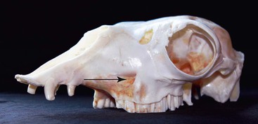
The infraorbital nerve (the rostral progression of the maxillary nerve) is blocked as it exits the infraorbital foramen, which is identified as a depression 1 to 3 centimeters (cm) (depending on the size of the camelid) directly above the premolars (Figure 48-2). The needle should be inserted into the canal, and 1 to 2 milliliters (mL) (depending on the size of the camelid) of local anesthetic should be injected. The tissues that are desensitized in small animals includes the ipsilateral maxilla and intraoral soft tissues; dentition; and extraoral soft tissues, including nose, upper lip, and skin from the site of the infraorbital foramen cranial to midline.4 It is assumed that the same structures would be desensitized in camelids.
Mandibular or Alveolar Block
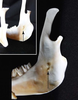
The mandibular nerve enters the bony cavity of the ramus of the mandible at the mandibular foramen, which is located directly dorsal to the most prominent portion of the most caudoventral aspect of the mandible immediately rostral to the ventral curve of the ramus (Figure 48-3).5 The nerve courses in the bony cavity until it exits at the mental foramen (described in the next section). Thus, the nerve is inaccessible once it has entered the bony cavity, and blockade must be performed at either the mandibular foramen or the mental foramen.

Figure 48-3 The mandibular foramen is indicated by the arrow. The direction that the arrow is pointing is the direction that the needle should be inserted when blocking the mandibular nerve. The inset photo emphasizes the location of the foramen on the lingual side of the mandible.
The mandibular foramen is approached extraorally by inserting a 1.5-inch (or longer) needle perpendicular to the ventral border of the mandible at the point just described. The needle should be kept close to the bone (it should be felt “scraping” the bone) so that it does not exit the oral mucosa, which would result in deposition of the local anesthetic into the oral cavity rather than over the mandibular nerve. In dogs and cats, a finger may be placed intraorally over the site of the block, and the exact location of the needle can be determined. This is difficult in camelids, and the block is made simply by determining the correct landmarks. Once the needle is completely inserted, 1 to 3 mL of local anesthetic should be injected. In dogs and cats, this block will provide analgesia of the dentition, and both the intraoral and extraoral soft tissue structures from the site of the mandibular foramen to the midline.4 The same pattern of blockade is expected in camelids. Because mandibular dental surgery in camelids is fairly common, this block is used routinely. As stated at the beginning of this chapter, local blocks may be used to improve anesthetic safety by allowing a decrease in the dose of maintenance anesthetic drugs in all species, including camelids.5 The block has been described as part of an analgesic multimodal protocol in an alpaca with dental disease.6
Stay updated, free articles. Join our Telegram channel

Full access? Get Clinical Tree


