Fluid Therapy


Fluid Therapy


Indications


Fluid therapy is an important part of the treatment plan for most sick camelids. As a rule of thumb, when camelids are recognized to be ill, they often have moderately to severely advanced disease. They are often dehydrated to some degree and may be azotemic and have electrolyte or acid–base imbalances as well. Camelids recognized as being ill often respond best to an aggressive treatment approach, which includes supplemental fluids. That being said, the volume of fluids given under these circumstances need not always be “aggressive.” In many cases, modest volumes of fluids evoke the desired response, and greater volumes are unnecessary and potentially detrimental.


The goals of fluid therapy in camelids are similar to those in other species. The major indications are to correct dehydration; to increase cardiac output, arterial blood pressure, and the perfusion of various organs; to correct electrolyte, mineral, and acid–base imbalances; to affect diuresis and increase urination; to provide a vehicle for other treatments; rarely to increase GI, pulmonary and other secretions; and ultimately to maintain a corrected patient at an acceptable level of the above functions.


Generally, three main indications exist for replacement fluids in camelids and a myriad of minor applications. The three major indications are (1) rapidly dehydrating conditions, where large-volume replacement may be indicated; (2) slowly dehydrating conditions, where replacement volumes are modest, but continued need is likely; and (3) conditions of vasculopathy or decreased cardiac output. Minor indications exclusive of these include whole blood or plasma transfusions to treat anemia or hypoproteinemia and the use of crystalloid fluids to administer or decrease the toxic effects of other medications.


Rapidly dehydrating conditions are the least common of the three main applications. Examples include acute C1 acidosis, diarrhea, loss of ingesta and secretions because of esophageal obstruction or toxic gastritis (rhododendron and related plants), and intestinal strangulations. A neonate that has never nursed could also fall into this category. These animals are characterized by having acute copious loss of water and usually electrolytes, without necessarily losing or catabolizing much protein in the process. Thus, they may have hyperalbuminemia or hyperproteinemia and packed cell volume (PCV) values at the higher end of the reference range.


Slowly dehydrating conditions are common and numerous. Thus, a variety of other abnormalities may accompany dehydration, and the animal may have adapted somewhat to compensate for the gradual fluid loss. Affected camelids frequently have low-grade anemia of chronic disease and evidence of protein loss or catabolism, and this may not have blood evidence of hemoconcentration.


Vasculopathies and processes that depress cardiac output without specifically causing dehydration are also relatively common. Examples include septic shock caused by neonatal bacterial infection, a ruptured ulcer, bacterial enteritis, or streptococcal peritonitis, heat stress, and ingestion of cardiotoxic compounds. Affected camelids may or may not be dehydrated but may display evidence of lack of adequate perfusion. These conditions may also complicate fluid therapy protocols because leaky vessels and poor heart function may negate some of the benefits of the additional intravascular volume.


Assessment


Determining the need for fluid therapy and formulating a plan for routes, rates, and components involve a combination of clinical intuition, physical examination findings, and laboratory analyses. Physical examination is most useful for finding signs of overt dehydration or subtler signs of poor peripheral perfusion. This may be complicated in camelids because, as a race, they appear to be well adapted to intermittent feed and water deprivation and may endure dehydration better compared with other domestic hoofstock species. Sunken, dull eyes, dry mucous membranes, and prolonged skin tent may all be found. Neck and eyelid skin appear to be the best places to assess skin tent time. Jugular fill is difficult to assess, but lack of filling after a long period of holding off the vein may provide some indication of severe dehydration. Heart rate may also increase, but it is important to note that dehydrated or adult camelids in shock frequently have heart rates ranging from 72 to 90 beats per minute (beats/min), which are within some published reference ranges. All heart rates greater than 72 beats/min should be considered potentially abnormal in adult camelids, as they may be a sign of dehydration, low cardiac output, or stress.


Behavioral evidence of dehydration or poor cardiac output may include water-seeking behavior, lethargy, inappetence, and decreased urination. Clinicopathologic evidence of dehydration includes hemoconcentration, evidenced by a high PCV, high blood protein or albumin concentrations, or both. It is crucial to keep in mind that these abnormalities are frequently masked by concurrent protein loss, catabolism, blood loss, anemia, or chronic disease and that refractometer readings of protein may be confounded and artificially high in camelids with hyperlipemia. Other laboratory evidence of dehydration includes the presence of azotemia, lactic acidosis, and electrolyte abnormalities. Azotemia may be masked or difficult to interpret in some cases; with acute inappetence, blood urea moves into the forestomach and is deaminated by gastric microbes. This effect lasts approximately 3 days, after which microbial die-off diminishes this catabolic pathway. Blood creatinine may also be low in camelids with chronically low muscle mass, as in neonates or chronic poor-doers. To avoid being confounded by one of these processes, measurement of both blood urea nitrogen (BUN) and creatinine concentrations is recommended. These confounders may also affect the rule of thumb, that a BUN-to-creatinine ratio (both in milligrams per deciliter [mg/dL]) of 20 : 1 or greater reflects prerenal azotemia, so this interpretation should be made with some caution.


Lactic acidosis may reflect global (hypovolemia, poor cardiac output) or focal (strangulation, thrombosis) anaerobic metabolism. Both are indications for fluid therapy and may be caused by overt dehydration or other forms of shock. Electrolyte abnormalities are suggestive of pathologic conditions associated with dehydration and thus may be helpful. Hypernatremia is common in camelids and usually reflects loss of water from hyperglycemia and glucose diuresis. Moderate to severe hypochloremia, especially when accompanied by metabolic alkalosis, often reflects a small intestinal obstruction with decreased gastrointestinal absorption and pooling of water in the intestinal tract. Hyponatremia, especially when accompanied by hypochloremia and metabolic acidosis, often indicates loss of fluid and electrolyte through the digestive tract, especially when it is caused by enteritis or esophageal obstruction. Decreased pulse strength, poor mucous membrane color, and slow capillary refill time may all indicate dehydration or poor cardiac output. These may be further confirmed by diagnostic tests, including measurement of central venous pressure or cardiac function.


In addition to the difficulty in recognizing the need for fluid therapy, a number of other factors must be considered. Sick camelids are often fragile. They may not be as tolerant of procedures as healthy camelids would be, and undergoing too much stress may cause a rapid decline in their condition. Catheterization attempts should be carefully planned for success, other invasive or semi-invasive treatments or diagnostic tests should be performed gradually or delayed until the patient has had some time to recover, and some invasive interventions should not be attempted at all. Also, the need for fluids and concerns of edema are closely related. The high prevalence of hypoalbuminemia (61% of our hospital population of camelids) and hypoproteinemia (35%) in sick camelids dictates conservative rates of fluid administration or use of a colloid solution. Even normoproteinemic camelids frequently develop hypoproteinemia during hospitalization, so monitoring this aspect of the patient must be an ongoing process. In addition to dictating the frequent use of colloids, camelid physiology and pathophysiology affect the value of other components of fluids. Sick camelids are often hyperglycemic and assimilate exogenous glucose poorly, so it should not be given without indication, and concurrent use of insulin or insulinotropic medications should be considered. Lactate is also likely to accumulate in sick camelids, so it should likewise be avoided in fluids, when possible.


Oral Fluids and Orogastric Intubation


Allowing a thirsty camelid to drink water is the simplest way to provide oral fluids. Overhydration by this route is extremely uncommon, and polydipsia is rarely encountered. The amount that can be offered at one time is unknown. It is likely that water flows aborad relatively quickly in camelids with good gastrointestinal function, but this cannot be ensured in sick camelids. Electrolyte solutions may be used in place of water if replacement is necessary, but the high prevalence of hypernatremia in sick camelids mandates that these solutions not be used without evidence of need.


If the thirst response is inadequate, which is regrettably the case in many sick camelids, oral fluids are best administered via an orogastric tube. Small diameter tubes may also be passed through the nose, but camelids have narrow nasal passages and often resist this procedure more than the passing of a tube through the mouth. However, in crias, small-bore feeding tubes may be passed nasally and left in place.


Passing an orogastric tube in an adult camelid may induce struggling and a stress response, so it is important to assess whether the patient can tolerate the procedure, to make a plan to perform the procedure quickly and competently, and to abort the procedure, if necessary. Alpacas may be manually restrained, whereas llamas may be better restrained in a llama chute. Light sedation may help, for example, with butorphanol (0.022 to 0.05 milligrams per kilogram [mg/kg], intramuscularly [IM]), which has fewer deleterious effects on airway protective responses or cardiac output compared with many other sedatives. To prevent damage to the tube because of chewing by adolescents or adults, a piece of PVC pipe or a block of wood with the edges rounded and a hole drilled through the center for the tube should be used as a speculum (Figure 32-1); crias may be tubed without a speculum. When a block speculum is used, it is introduced from the side of the mouth into the interdental space and seated on the molars. The tube is passed through the hole, over the base of the tongue, and into the esophagus. Negative pressure when sucking back on the tube, palpating the tube adjacent to the cervical trachea, or hearing fluid bubbles over the abdomen when blowing on the tube confirm correct placement. The camelid may respond by struggling, regurgitating, or going into respiratory distress. Any of these may lead to reassessment of the adequacy of the restraint or the safety of the procedure and potentially cancelling or delaying it.
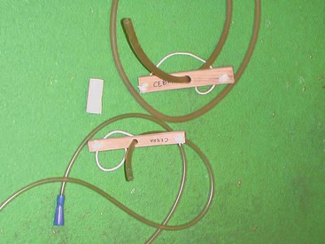

Stay updated, free articles. Join our Telegram channel

Mar 27, 2017 | Posted by in GENERAL | Comments Off on Fluid Therapy

Full access? Get Clinical Tree

Get Clinical Tree app for offline access