M. Kimberly J. McGurrin
Investigation of Cardiac Arrhythmias
Arrhythmia is any abnormality in the normal sequence of electrical activation in the myocardium. The normal front of depolarization is initiated at the sinus node, is conducted through the atria to the atrioventricular node, then travels into the His bundle and the Purkinje fiber system to depolarize the ventricles in a coordinated manner. The horse has an extensively arborized Purkinje system, which enables essentially simultaneous depolarization of the ventricles. The effect of abnormal conduction depends on the site of aberrant impulse generation, frequency of aberrant impulses, whether aberrant impulses occur in runs, and any impacts on overall heart rate.
Given that horses are primarily considered to be athletes, arrhythmias are often a cause for concern because of the potential impact on performance. Certain observations should prompt further investigation as a result of suspicion of an arrhythmia. The horse may present with an irregularity in the cardiac rhythm at rest and have profound clinical signs or poor performance. Arrhythmias, whether single ectopic beats or runs, have been identified in poorly performing horses at exercise and have been considered contributory if not causal of poor performance.
A complicating factor in the investigation of the poorly performing horse is that benign dysrhythmia is common. On careful examination by auscultation and review of electrocardiogram (ECG) recordings, as many as 25% of horses have been reported to have dysrhythmia at rest. The horse’s heart at rest is subject to a high level of parasympathetic control, more so than other domestic species. As a result, there is a predisposition to vagally mediated dysrhythmias. On auscultation, these rhythms sound abnormal. However, they are generally considered to be physiologic, have no underlying pathology, and have no influence on performance. With reductions in vagal tone, such as excitation or exercise, these arrhythmias abate.
It is usually more difficult to determine the significance of an arrhythmia with certainty when it does not appear to be vagally mediated or fails to manifest when the horse is at rest. In these cases, a comprehensive investigation of cardiac dysrhythmia is needed.
Physical Examination
There are no diagnostic tools that can replace a careful physical examination. Pulse palpation and cardiac auscultation are the initial tools that identify an arrhythmia. It is by the use of these skills that one determines whether an arrhythmia is present at rest, and whether it is present at all times during rest or is intermittent in nature. By auscultation, one may also determine the effects of the arrhythmia on heart rate and overall rhythm. Given that subtle variations in cardiac rhythm are normal, perfect rhythm indicates a complete absence of autonomic input. With auscultation alone, the examiner can determine the effects of light exercise on horses with nonclinical bradydysrhythmias, infrequent pauses, or ectopic beats.
Resting Electrocardiogram
A resting ECG should be performed in any horse in which an arrhythmia is suspected. It is the most important diagnostic tool when a cardiac rhythm is abnormal at rest, and is invaluable in identifying potentially life-threatening dysrhythmias. When intermittent dysrhythmia or dysrhythmia at exercise is suspected, a baseline resting ECG is warranted to establish the horse’s “normal” sinus rhythm.
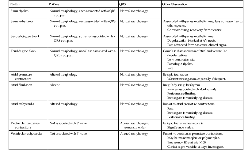
In an animal that has no evidence of cardiac disease and no history of poor performance, a resting ECG alone is adequate. In an animal with a history of poor performance or clinical signs suggesting dysrhythmia, a resting ECG alone may provide insufficient information. Many performance-limiting dysrhythmias do not manifest at rest (atrial fibrillation is an important exception), and detection of intermittent arrhythmias may necessitate prolonged recording periods. Electrocardiogram findings in common arrhythmias are summarized (Table 121-1).
In humans and small animals, a multiple-lead ECG may be used to generate information about chamber enlargement; normal values have been well established. However, in the horse, a multiple-lead ECG is of less value because of the extensive branching of the Purkinje system in the ventricular wall. A single lead system is normally sufficient. A base–apex or similar system is most common. The goal in placing the electrodes is to optimize the signal along the direction of the maximal deflection of the electrical impulse generated within the ventricle. To do this, the electrodes are placed essentially parallel to this impulse.
A common and satisfactory option for arranging the electrode system starts with placing the negative electrode labeled RA (right arm; often white) at the manubrium. The positive electrode labelled LA (left arm; black) can be placed at the xiphoid. The ground electrode labeled LL (left leg; green or red) may be attached to either side of the neck. Consistency in lead positioning facilitates interpretation. With this configuration, the ECG machine should be set to lead 1 (the other ECG control settings vary the polarity of the limb leads). A paper speed of 25 mm/second is standard for equine ECG recordings. A faster speed is sometimes used to capture greater detail. It is important to ensure that the horse is well restrained and standing still to optimize the recording. It is also important to run the ECG for long enough to obtain adequate sample recording. The horse has a low resting heart rate, and when evaluating for an arrhythmia, it is important to collect enough recording to enable assessment of the cardiac rhythm. For example, 15 seconds of recording at a heart rate of 30 beats/minute is less than 8 beats. Recordings of 30 seconds or longer may be required for diagnostic accuracy.
Interpretation of the Resting Electrocardiogram
To reliably interpret an ECG recording, one should understand the generation of cardiac electrical impulses and the variations in normal waveforms. The analysis should be approached in a standardized manner to minimize error and improve repeatability. To ensure the ECG recording is of diagnostic quality, the examiner should evaluate it for artifacts, such as irregularities caused by movement or electrical interference. Artifacts caused by movement or poor electrode attachment generally appear as coarse, variably sized, irregular variations on the ECG. The key in differentiating artifacts from ectopic impulses is that artifacts are not followed by T waves. Electrical interference tends to cause fine, high-frequency variations in the signal at much too high a frequency to be cardiac in origin.
Next, the examiner moves on to sequentially assess heart rate, heart rhythm, and any abnormal complexes that may be present. A bifid (two deflections) P wave is common in the horse and is associated with asynchronous depolarization of the atria, with the right atrium depolarizing first. Biphasic and single positive deflections are also normal variations. P wave morphology may also change without alteration in cardiac rhythm because of variation in the direction of the wavefront of depolarization. Prominent downward deviation in the PR segment is a result of atrial repolarization (Ta wave).
Normal conduction through the AV node is slow and is seen on the ECG recording as the delay between the P wave and QRS complex (up to 0.5 second in duration). The first positive deflection in the QRS complex is the R wave. However, the horse does not commonly have an R wave in the base–apex lead system, so the main deflection observed is downward, essentially a large S wave. T waves are relatively labile in morphology among horses and can vary with heart rate within an individual horse. It is often difficult to detect the end of the T wave.
Stay updated, free articles. Join our Telegram channel

Full access? Get Clinical Tree


