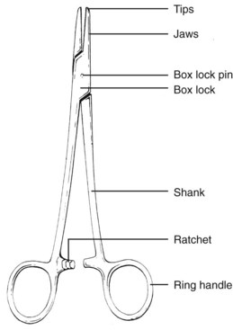
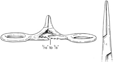
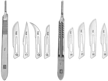
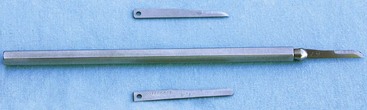
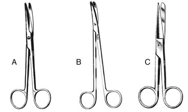
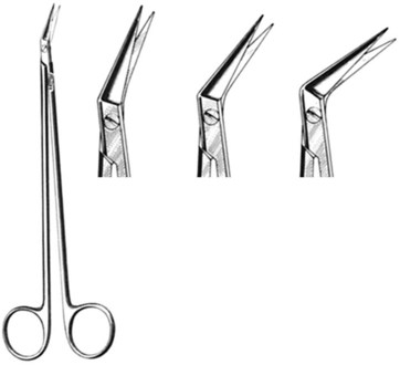
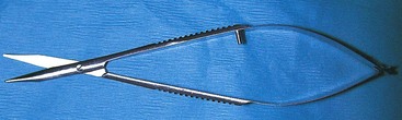
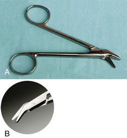
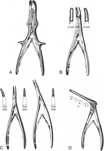
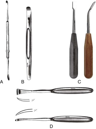
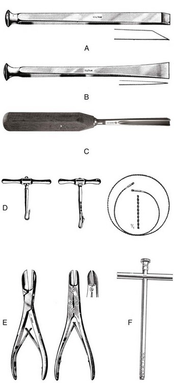
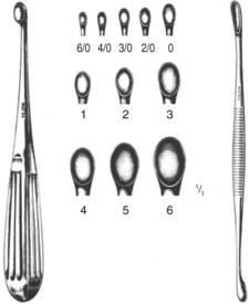
Chapter 13 Surgical instruments are defined as hand-held tools or implements used by health professionals for the performance of surgical tasks.5 Surgery always has been intimately connected with its instruments.13 Instruments both serve the surgeon and influence how procedures can be conducted.13 An essential requirement of any instrument is the ability to complete the intended task in an efficient and precise manner.21 The basic parts of an articulating instrument are shown in Figure 13-1. These parts include the tips, jaws, box lock and pin, shank, ratchet, and ring handles. The tips of many instruments (e.g., scissors) are designed for specific uses. Tips of grasping instruments may be straight or curved, with the latter tip configuration generally aiding visibility. Instruments intended to grasp tissue have jaws designed to be traumatic or atraumatic with various configurations and combinations of serrations and teeth. Instruments with jaws that have prominent serrations or coarse teeth are usually more traumatic than those with more subtle serrations or finer teeth. The box lock and pin is the joint or hinge of the instrument, and, as such, it absorbs stress during instrument preparation (autoclaving) and use.17 Failure of the box lock of an articulating instrument is a relatively frequent reason for limited instrument useful life. The shank determines the overall length of the instrument, which should match both the surgeon’s hand and the intended use. Instruments with longer shanks tend to be used to access tissues within body cavities. The ratchet has interlocking teeth to keep an instrument jaw in the closed position (Figure 13-2).17 As such, the ratchet helps to stabilize the instrument and either the tissue (e.g., blood vessel) or the object (e.g., suture needle). Ring handles serve as a means of using and controlling the instrument.17 Proper positioning of digits within the ring handles is an important determinant of surgical effectiveness and efficiency (see Chapter 19). This chapter includes descriptions of commonly used surgical instruments, instrument care, and instrument problem solving. The following instruments are described: cutting instruments, including scalpel, scissors, rongeurs, and periosteal elevators; bone-cutting instruments, including chisels, osteotomes, gouges, saws, bone-cutting forceps, curettes, and trephines; grasping instruments, including needle holders, crushing and noncrushing tissue forceps, hemostatic forceps, thumb forceps, towel clamps, and bone-holding forceps; retractors, hand-held and self-retaining; and suction tips, miscellaneous instruments, and microsurgical instruments. Instrument handling and use is covered in Chapter 19. The scalpel is one of the primary cutting instruments of the surgeon.12 Most scalpels are handles that accept disposable blades. Such blades are made of carbon steel or stainless steel and are designed to fit standard #3 or #4 handles. The #3 scalpel handle, which is used primarily in small-animal surgery, accepts blades #10, #11, #12, and #15; the #4 handle accepts blades #20 to #25 (Figure 13-3). Each handle is spatula-like, has ribbed grip areas, and may have a measurement scale.19 A long version of the #3 handle is available for working deep in the thoracic cavity. A specialized rounded Beaver scalpel handle is used in ophthalmic surgery and accepts Beaver blades of various shapes (Figure 13-4).11 In large part because of the sharpness of the cutting edge, scalpel incisions produce less tissue trauma than do scissor incisions. Scissors are available in various shapes, lengths, and weights. Scissors may be classified by their point type (sharp-sharp, sharp-blunt, and blunt-blunt), blade shape (straight or curved), and cutting edge type (plain or serrated). Scissors with straight tips have a greater mechanical advantage; those with curved tips have greater versatility and enable improved visibility, particularly deeper in wounds.22 Serrated cutting edges help prevent slippage, particularly on lax (e.g., eyelid) or dense (e.g., cartilage) tissue.22 Scissors used commonly in small-animal surgery include Metzenbaum, Mayo, and utility or operating scissors (Figure 13-5).17 Metzenbaum scissors have thin delicate blades that are approximately one fourth of the overall instrument length. They are used on relatively delicate tissue (e.g., subcutaneous tissue). Mayo scissors have thick blades that are approximately one third of its length. They are used on denser tissue (e.g., fascia). Utility or operating scissors often have straight blades with sharp-blunt tips. They are often reserved for use on inanimate objects (e.g., suture material). Specialized scissors include Martin cartilage scissors, which have coarser blades with serrated edges for cutting denser tissues (e.g., during otic procedures); Potts-Smith scissors (Figure 13-6), with various angled blades for cardiovascular procedures; and tenotomy scissors, which may be ring-handled (e.g., Stevens) or spring-handled (e.g., Wescott) for ophthalmic procedures (Figure 13-7). Wire scissors frequently have short, notched serrated blades and are used to cut stainless steel wire, usually during orthopedic procedures (Figure 13-8).19 Rongeurs are forceps with cupped jaws and blunted or tapered tips.12 They are used in neurosurgical and orthopedic procedures to remove pieces of bone to expose underlying structures, to prepare bone for grafting, or to recontour bone ends.14 The jaws may be straight, curved, or angled, and the mechanism for closure may be single or double action.14 Double-action rongeurs have a greater mechanical advantage and require less force during use.12 Selection of appropriate rongeur type varies with the density of the bone to be removed (e.g., spinal column vs. tympanic bulla), the proximity of adjacent structures (e.g., spinal cord), and the degree of surgical exposure (e.g., long bone fracture repair vs. neurosurgical decompression).14 Examples include the Stille-Luer and Ruskin, both double-action rongeurs (used on dense bone in well exposed areas [e.g., recontouring long bone ends]); Lempert, which has finer, more delicate jaws (used on less dense bone in more restricted areas [e.g., bulla osteotomy]); and Kerrison, which has a single cutting blade and a footplate (used during ventral slot neurosurgical decompressive procedures) (Figure 13-9). Periosteal elevators are available in various shapes and sizes, have a round or straight edge, and are used during many orthopedic and neurologic, as well as selected soft tissue, surgeries.17 Periosteal elevators are helpful in reflecting muscle and other soft tissue from bone.14 Examples include the Freer and Sayre, both double-ended elevators, and ASIF (Synthes/ AO) and Langenbeck single-ended elevators (Figure 13-10). Bone-cutting instruments include chisels, osteotomes, gouges, saws (manual and power), bone-cutting forceps, curettes, and trephines (Figure 13-11). A chisel is beveled on one side, and osteotomes are double beveled; each is used most frequently with a mallet. Gouges and manual saws are used less commonly and are used to reshape or cut bone. Manual saws used in veterinary surgery include the hobby saw and the Gigli wire saw. Power (oscillating) saws are used commonly for various orthopedic (e.g., tibial plateau leveling osteotomy or femoral head and neck ostectomy) and soft tissue procedures (e.g., median sternotomy). Such saws are often used with procedure-specific blades (e.g., tibial plateau leveling osteotomy, sagittal). Bone-cutting forceps come in various sizes and weights, and, similar to rongeurs, are available as single or double action; they have paired chisel-like tips for cutting bone. Curettes, available in various sizes and shapes, including right-angle, have a cupped end that is used to remove bone or soft tissue, particularly from restricted sites (e.g., cancellous bone harvesting, bulla osteotomy) (Figure 13-12). The cupped end is sized by using terminology similar to but not corresponding with that used for suture material. Trephines have t-handles, a cylindrical cutting blade, and often a central stylet for removing bone from the shaft.12 Trephines have been used to obtain samples of bone for biopsy and to gain initial entry into bony cavities. Most needle holders are equipped with a ratchet, and some have suture-cutting ability.9 Needle holders are available in various sizes and lengths. The jaws of the needle holder are short and frequently have grooves that are cross-hatched on the surface.19 Such jaw designs (e.g., cross-hatching) are intended to limit twisting and rotation of the needle, allowing the surgeon to accurately control passage of the needle through tissue.4 Needle-holder jaws should match both the size and the type of needle being used. Textured tungsten carbide needle-holder jaw inserts have advantages over both smooth jaw inserts and those with teeth.4 Such inserts provide intermediate needle-holding security between smooth needle-holder jaws and those with teeth, while minimizing the potential for suture material damage and weakening during knot tying.4 Considerations when selecting the appropriate needle holder include the magnitude of force required to overcome the ratchet mechanism and the design of the jaw.3 The angle of each tooth of the ratchet mechanism should be 39 degrees rather than 45 degrees to enhance the security of the engagement of the interlocking teeth.4 Ideally, the surgeon should use a needle holder with a clamping moment less than that of the yield moment of the needle.4 Needle holders used frequently in veterinary surgery include Mayo-Hegar and Olsen-Hegar (Figure 13-13), a combination needle holder and scissors. In large part because of the combined functionality of the Olsen-Hegar needle holder, it tends to have a shorter useful life than other needle holders. Some ophthalmic needle holders (e.g., Castroviejo) are equipped with spring handles that catch or release with gentle pressure (Figure 13-14).19
Instrumentation
Surgical Instruments
Scalpel
Scissors
Rongeurs
Periosteal Elevators
Bone-Cutting Instruments
Grasping Instruments
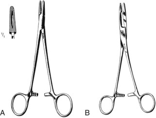
Needle Holders
< div class='tao-gold-member'>
![]()
Stay updated, free articles. Join our Telegram channel

Full access? Get Clinical Tree


Instrumentation
Only gold members can continue reading. Log In or Register to continue
