Fig. 1
ASFV viral DNA digested with restriction enzyme. (a) Lane 1: DNA ladder. Lane 2: two microliters (400 ng) of non-digested ASFV viral DNA. (b) Lane 1: DNA ladder. Lane 2: ten microliters (763 ng) of MboI digested ASFV viral DNA
4.
Label the DNA fragments using a DNA labeling system, following the instructions of the fabricant (see Note 14 ).
5.
After DNA labeling, purify probes using a DNA purification kit and quantify the concentration of the labeled DNA (see Note 12 ).
6.
Mix 300 ng of DNA-labeled probes (see Note 15 ) with 30 μg of competitor DNA and add water to obtain a precipitable volume (minimum volume of 30 μl).
8.
3.2 In Situ Hybridization
2.
Rehydrate the tissue sections through an ethanol series (2 times in absolute ethanol, twice in 96 % ethanol, and once in water, 3 min each time) and wash in 1× automation buffer (see Note 20 ).
3.
4.
After the pretreatment with protease solution, wash all sections with 1× automation buffer.
5.
6.
7.
Following tissue sections denaturation, distribute the 33 μl of each DNA probe preparation over each section, cover them with coverslips, seal with rubber cement, and incubate in a humidified box at 37 °C overnight (see Note 25 ).
8.
Following the overnight hybridization step, immerse the preparations in 2× SSC to allow the coverslips to drop off.
9.
Wash the slides 2× with wash solution 1, 5 min each time.
10.
Wash the slides with wash solution 2 at 37 °C for 5 min.
13.
Rinse the slides 2–3 times with blocking solution.
14.
Rinse the slides 3–4 times with 1× automation buffer.
15.
Wash the slides with wash solution 3 for 5 min.
16.
17.
Rinse the slides 1–2 times with buffer 3 to stop the color reaction and deep the preparations into buffer 3 until the Fast Green counterstaining step.
18.
Counterstain the preparations with Fast Green solution for 10 min.
19.
Wash with distilled water.
20.
Get Clinical Tree app for offline access

Mount sections and examine by light microscopy (Fig. 2) (see Notes 30 and 31 ).
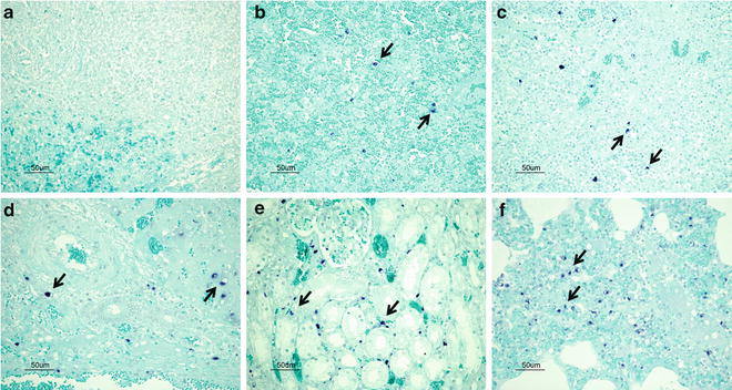
Fig. 2


ASFV-DNA detection using DIG-labeled probes in formalin-fixed paraffin-embedded tissues. (a) Negative lymph node. (b–f) Positive tissues, where dark-blue dots in the preparation correspond to the presence of viral nucleic acids (examples of positive labeling are indicated with arrows). (b) Spleen. Intense positive detection in red pulp macrophages and trabecular medullary areas. (c) Lymph node. Moderate positive detection in perifollicular macrophages (marginal zone). (d) Liver. Moderate to intense positive detection in sinusoidal and alveolar macrophages and Kupffer cells. (e) Kidney. Intense positive detection in circulating macrophages and renal endothelium. (f) Lung. Intense positive detection in pulmonary alveolar, vascular, and parenchymal macrophages
Stay updated, free articles. Join our Telegram channel
Full access? Get Clinical Tree