51 Hypertensive retinopathy
CLINICAL EXAMINATION
Ophthalmic examination can be similarly variable. One or both eyes can be affected. Some patients will be blind, with no menace response and dilated poorly responsive pupils. Remember that cats can retain a brisk pupillary light reflex despite advanced retinal disease, so these should not be relied upon in this species. Dazzle reflexes are usually reduced. The conjunctiva is occasionally slightly hyperaemic but no ocular discharge or corneal ulceration is usually present. Some hyphaema or vitreal haemorrhage might be present, both of which render fundus examination impossible (see Figure 36.2). However, it is unusual for both eyes to have severe haemorrhage simultaneously and so some fundus evaluation should be possible.
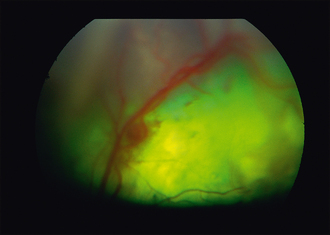
If any opacification of the ocular media is present, indirect ophthalmoscopy will be more rewarding than direct ophthalmoscopy. The retina might be completely detached in one or both eyes, and can be seen as a grey veil floating behind the lens (see Figure 49.1). In less advanced cases, the fundus will show various changes – some small blister-like areas of bullous detachment (seen as elevated, out-of-focus dull patches within the tapetal fundus) or retinal haemorrhages – both within the retina itself (flame shaped) and pre-retinal (dark puddle-like areas). The retinal blood vessels are frequently engorged and tortuous with some perivascular cuffing and haemorrhage (Figure 51.1). The vessels often have variable diameter along their length, giving the appearance of a string of sausages. Variable amounts of vitreal haemorrhage can also be present. Although bilateral, the condition is not symmetrical.
CASE WORK-UP
Routine haematology and biochemistry is essential, together with appropriate tests for hyperthyroidism in cats, but also sometimes hypothyroidism in dogs. Blood pressure should be measured as outlined in Table 51.1. Normal readings are provided in Table 51.2.
Table 51.1 Measurement of blood pressure in dogs and cats


