Gastrointestinal, Pancreatic, and Hepatic Disorders

9 Gastrointestinal, Pancreatic, and Hepatic Disorders


Some gastrointestinal (GI) problems (e.g., vomiting, diarrhea, weight loss, anorexia, icterus, hepatomegaly, abnormal behavior associated with eating, abdominal pain) typically necessitate laboratory testing. On the other hand, dysphagia, regurgitation, ptyalism, halitosis, constipation, mucoid stools, hematochezia, and melena are usually best approached initially by other means (e.g., physical examination, radiology, ultrasonography, endoscopy, laparotomy, and/or biopsy).


Differentiation of Expectoration, Regurgitation, and Vomiting


Whenever fluid, mucus, foam, food, or blood is expelled from the mouth, one must determine whether vomiting, regurgitation, gagging, or expectoration is occurring. The history sometimes allows differentiation.


Regurgitation


Regurgitation is due to oral, pharyngeal, or esophageal dysfunction and is typically characterized as a relatively passive expulsion of esophageal contents. Gagging is the expulsion of oral or pharyngeal material and may be associated with disorders causing dysphagia (i.e., difficult swallowing) or regurgitation. The relatively minor abdominal contractions associated with gagging are typically different than the vigorous abdominal contractions that classically occur with vomiting. Regurgitation may follow seconds to hours after eating or drinking. Patients may regurgitate white foam (i.e., salivary secretions that have been swallowed) and/or food. Regurgitated food material is undigested and sometimes has a tubular form conforming to the shape of the esophageal lumen. Most clients cannot reliably distinguish undigested from digested food. Regurgitated material that has remained in the esophagus for long time periods can appear “partially digested” because it is macerated, odoriferous, and mixed with saliva. If blood is present, it is usually undigested (i.e., bright red), whereas blood originating from the stomach is usually partially digested by gastric acid and has a “coffee grounds” appearance readily distinguishing it from the undigested form (unless the patient vomits before the blood can be partially digested).


It is sometimes difficult to differentiate vomiting from regurgitation via history, and in some patients the processes are concurrent. Vomiting may cause secondary esophagitis and subsequent regurgitation, or a patient with long-standing esophageal disease may develop another concurrent disorder causing vomiting. It is therefore important to clarify the chronologic order of specific signs. Finally, some patients with signs “classic” for regurgitation are vomiting instead. To aid in differentiation, one may attempt to observe the act of expulsion by feeding the patient, although this is very unreliable (“watched” regurgitating patients often do not regurgitate). Watching the patient eat occasionally helps if there is obvious pharyngeal dysphagia that suggests oropharyngeal disease. Some patients with pharyngeal dysphagia also have concurrent esophageal dysfunction. Contrast radiographs and/or fluoroscopy of the pharynx and esophagus can usually differentiate vomiting from regurgitation.


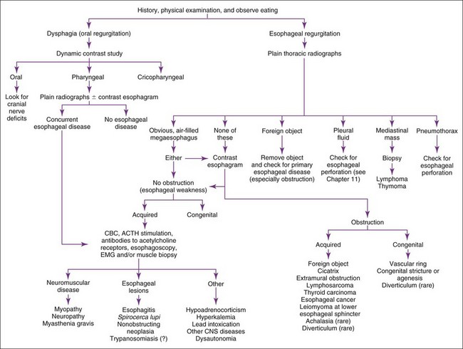
Regurgitation is usually best evaluated by history, physical examination, plain and contrast radiographs, and/or esophagoscopy (Figure 9-1). Contrast radiographs should use barium instead of iodide contrast agents unless esophageal rupture is suspected (e.g., finding air or fluid in the mediastinum on plain radiographs). The main purpose of a contrast esophagram is to distinguish esophageal motility abnormalities from anatomic lesions (e.g., obstruction, mass, inflammation, fistula). Some drugs (e.g., xylazine, ketamine) can cause esophageal hypomotility, making the radiographs potentially misleading. Esophagoscopy is insensitive for diagnosing esophageal muscular weakness but sensitive for finding anatomic lesions, differentiating intramural from extramural obstruction, identifying esophagitis, and removing foreign objects. Patients with acquired esophageal weakness should be evaluated for myopathies, neuropathies, and myasthenia gravis (generalized or localized to the esophagus). Occasionally, hypoadrenocorticism, hyperkalemia, lead poisoning, Spirocerca lupi, and selected central nervous system (CNS) disorders (e.g., distemper, hydrocephalus) may be responsible. Generalized or localized myopathies and neuropathies have several causes (e.g., trauma, dermatomyositis, thymoma, botulism, tick paralysis, systemic lupus erythematosus, nutritional factors, toxoplasmosis, trypanosomiasis). Dysautonomia occurs in dogs and cats, causing generalized dysfunction of the autonomic nervous system producing esophageal hypomotility. It is important to detect underlying disorders so that one may treat the cause rather than just the symptoms. It is also wise to evaluate patients with unexpected esophageal foreign objects (e.g., a relatively small bolus of food) for partial obstructions (e.g., subclinical vascular ring anomaly, stricture).


Vomiting


Vomiting is a reflex act originating in the CNS that can be stimulated by various conditions. One must consider primary GI disease and non-GI disorders (e.g., metabolic, inflammatory, and toxic conditions) as causes of vomiting. Many vomiting patients have non-GI problems.


Vomiting is classically characterized by prodromal nausea (i.e., salivation, licking of lips) followed by retching or forceful abdominal contractions. Vomiting may occur any time after eating or drinking (seconds to hours). A patient may vomit food, water, fresh blood, or mucus that is indistinguishable from regurgitated material. Bile, partially digested blood (i.e., “coffee grounds”), or expelled material with a pH of 5 or less strongly suggests vomiting as opposed to regurgitation. Vomited duodenal contents may have a pH greater than or equal to 7 and are usually positive for bile. A urine dipstick with a pH indicator is useful in making pH determinations. A patient that has “dry heaves” is typically vomiting as opposed to regurgitating.


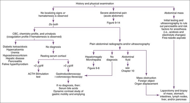
Vomiting patients are best divided into those with acute (<2 weeks) versus those with chronic (>2 weeks) vomiting. The most common categories of causes for each are listed in Boxes 9-1 and 9-2. Patients with acute vomiting often spontaneously resolve if they are supported by fluid therapy. A thorough history and physical examination are indicated first. Laboratory evaluation and/or imaging should be considered if the disease is severe or a serious disease (e.g., obstruction) is suspected. If vomiting persists, is progressive, or is attended by other clinical signs (e.g., polyuria-polydipsia [pu-pd], weight loss, icterus, painful abdomen, ascites, weakness, hematemesis), additional testing is indicated (Figure 9-2).


Pancreatitis


Acute pancreatitis occurs commonly. Predisposing causes in dogs include hyperlipidemia, fatty meals, or obesity. Pancreatitis can occur in any dog, but middle-aged obese female dogs, schnauzers, and Yorkshire terriers seem to be predisposed. Vomiting may or may not be associated with eating, abdominal pain, fasting hyperlipidemia, bloody diarrhea, and, rarely, diffuse subcutaneous fat necrosis. On radiographic examination, a mass or indistinctness due to localized peritonitis may be visible in the cranial right abdominal quadrant. CBC, serum amylase, and serum lipase activities are insensitive and nonspecific; patients with pancreatitis can have almost any result on these tests. Increased ALT and SAP concentrations (as the result of the proximity of the pancreas to the liver and obstruction of the biliary duct) are common but insensitive and nonspecific. Mild to moderate hypocalcemia sometimes occurs. Abdominal ultrasonography can be very specific for canine pancreatitis, but its sensitivity depends upon operator skill and timing (i.e., ultrasound findings can change dramatically in a matter of hours). If a pancreatic mass is discovered during surgery, it must be biopsied; chronic pancreatitis can be grossly indistinguishable from pancreatic neoplasia. The canine immunoreactive pancreatic lipase (spec cPL) (IDEXX, Westbrook, ME) test is the most sensitive test for pancreatitis, but its specificity for clinically important disease is still being determined. Chronic pancreatitis may be presumptively diagnosed if one finds exocrine pancreatic insufficiency in a breed that is not affected by pancreatic acinar cell atrophy.


Pancreatitis is an important but difficult-to-diagnose disease in cats. Chronic pancreatitis in older cats sometimes occurs in conjunction with cholangiohepatitis and/or inflammatory bowel disease (IBD) (often referred to as a triaditis syndrome involving all three organs). Vomiting is not as prominent in feline pancreatitis as it is in canine pancreatitis. Feline trypsin-like immunoreactivity (fTLI) concentrations are increased in some patients. Abdominal ultrasonography is specific, but the sensitivity is uncertain. A pancreatic biopsy may be required for a definitive diagnosis. The feline immunoreactive pancreatic lipase (spec fPL) test appears to be useful in diagnosing pancreatitis. Feline pancreatitis occasionally is due to toxoplasmosis or to feline infectious peritonitis (FIP) (see Chapter 15).


Abdominal Inflammation


Septic or nonseptic peritonitis (or inflammation of any abdominal organ) may cause vomiting. Abdominocentesis or abdominal lavage (see Chapter 10) may be needed, especially if physical examination or abdominal imaging suggests abdominal fluid. Occult cases may require laparoscopy or exploratory surgery for diagnosis.


Canine Immunoreactive Pancreatic Lipase (Spec cPL)


Gastrin


Acute Diarrhea


Patients with diarrhea are best classified into those with acute (<2 to 3 weeks) versus those with chronic (>2 to 3 weeks) diarrhea. Acute diarrhea (Box 9-4) is usually self-limiting, although some conditions may be severe and cause mortality (e.g., acute hemorrhagic gastroenteritis, parvoviral disease, hookworms, intoxication). History should explore the possibility of recent dietary change and exposure to infectious agents. Diet, bacteria, viruses, and parasites are the major identifiable causes of acute diarrhea in dogs and cats. Because intestinal parasites may contribute to any diarrheic state, fecal examinations (direct and flotation) are typically warranted in diarrheic patients. Giardiasis may require special diagnostic techniques (see Fecal Giardia Detection later in this chapter). The need for diagnostics depends upon (1) the severity of the problem (i.e., more severely ill patients require more diagnostics), and (2) the likelihood that the patient has an infectious agent that has potential to be nosocomial or zoonotic.


Feeding bland or elimination diets may be diagnostic and therapeutic. Depressed, weak, and dehydrated patients should undergo electrolyte and acid-base evaluations to aid in selecting fluid replacement therapy. All patients less than 12 to 14 weeks of age and those that are emaciated or weighing less than 3 kg should undergo blood glucose monitoring to detect secondary hypoglycemia. CBC is indicated in most febrile or severely depressed patients so that sepsis or transmural inflammation can be detected. Fecal cultures, ELISA, and/or polymerase chain reaction (PCR) for Salmonella spp., Campylobacter jejuni, Yersinia enterocolitica, verotoxin-positive Escherichia coli, Clostridium perfringens, or Clostridium difficile may be performed, but establishing a cause-and-effect relationship between the organism and disease can be difficult.


Not all patients with canine parvoviral diarrhea are severely ill, diarrheic, febrile, or have identifiable leukopenia. Leukopenia may only persist 24 to 36 hours and be missed if a CBC is not performed during that period. Other diseases causing severe sepsis (i.e., perforating linear foreign body with peritonitis or overwhelming salmonellosis) can cause leukopenia indistinguishable from that of canine parvoviral enteritis. Routinely used vaccination schedules do not necessarily guarantee protection against canine parvovirus. In-house ELISA tests for parvovirus performed on feces appear to be specific for parvoviral antigen, but testing may be negative if done too early or too late. Fecal shedding of viral particles may not occur for 1 to 3 days after signs begin and decreases rapidly with time. The test result should be strongly positive within 3 days of the onset of clinical signs and remain positive for several days. A recent vaccination may result in a weakly positive fecal ELISA.


Chronic Diarrhea


Chronic diarrhea should first be defined as either small intestinal or large intestinal in origin (Table 9-1). Occasionally, large and small intestines are concurrently involved. Patients with chronic diarrhea in which clinical disease is not severe are often treated with therapeutic trials before aggressive diagnostics are instituted. The specifics of the therapeutic trials are influenced by whether the patient has large or small bowel disease. Patients should usually have at least three fecal examinations at 48-hour intervals. If these tests are negative, it is still acceptable (depending upon the risk of parasites in the geographic location) to treat empirically for Giardia infection and whipworms before aggressive diagnostics are begun. Giardiasis may be particularly difficult to diagnose (see Fecal Giardia Detection later in this chapter). Adverse food reactions (i.e., allergy, intolerance, fiber deficiency) commonly cause chronic diarrhea. Dietary intolerances are a reaction to a particular substance in the diet, whereas true food allergies are immunologic reactions to specific antigens. Dietary food trials are indicated in suspected cases. There are antibiotic-responsive intestinal diseases that are also treated empirically; however, the specific therapy varies with whether the patient has large or small bowel disease (see next section). Failing to respond to empirical anthelmintic, dietary, and antibacterial therapy indicates the need for further diagnostics.


TABLE 9-1 DIFFERENTIATION OF CHRONIC SMALL INTESTINAL DIARRHEA FROM CHRONIC LARGE INTESTINAL DIARRHEA


  SMALL INTESTINAL DIARRHEA LARGE INTESTINAL DIARRHEA
Weight loss (very important criterion) Expected Uncommon except with severe disease (e.g., histoplasmosis, pythiosis, or cancer)
Polyphagia Often present Uncommon
Vomiting May occur Occurs in 10%–20% of patients
Volume of feces May be normal or larger than normal May be normal or smaller than normal
Frequency of defecation Normal to slightly increased Normal to markedly increased, may have many small defecations per bowel movement
Slate-gray feces (steatorrhea) Rare No
Hematochezia No Sometimes
Melena Rare No
Mucoid stools Rare (unless ileum is diseased) Often present
Tenesmus/dyschezia Rare Sometimes


Large Intestinal Disease


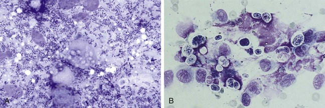
Large intestinal disease has different parasites (i.e., Trichuris vulpis, Tritrichomonas fetus), dietary problems (i.e., fiber-responsive diarrhea), and bacterial problems (i.e., so-called clostridial colitis that responds best to tylosin or amoxicillin) than small bowel disease. Once parasitic, dietary, and “clostridial colitis” are eliminated by diagnostics and therapeutic trials, additional diagnostic steps, such as rectal mucosal scrapings (not swabs) with cytologic examination (Figure 9-3) might be appropriate. Persistent large intestinal disease that fails to respond to these therapeutic trials or that is associated with hypoalbuminemia or obvious weight loss is usually an indication for abdominal ultrasound followed by fine-needle aspiration and/or colonoscopy-ileoscopy plus biopsy. Rigid colonoscopy of the descending colon is adequate for diagnosis in most cases. Flexible endoscopy allows access to the descending, transverse, and ascending colon; ileocolic valve; cecum; and ileum. If flexible endoscopy is unavailable, abdominal ultrasonography may reveal lesions in areas not accessible with rigid endoscopy.


Small Intestinal Disease


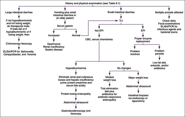
Small intestinal disease has different parasites (e.g., Giardia), dietary problems (e.g., lymphangiectasia), and bacterial problems (i.e., so-called antibiotic-responsive enteropathy [ARE] or dysbiosis that may respond to a variety of antibacterials) than large bowel disease. Chronic and severe small intestinal diarrhea necessitates differentiation of maldigestion, protein-losing enteropathy (PLE), and malabsorptive disease without protein loss (Figure 9-4). Weight loss and diarrhea are usually present, but some patients only have weight loss.


Maldigestion


Maldigestion due to bile acid insufficiency caused by biliary obstruction is rare. Intestinal lactase deficiency is uncommon, but a lactose-free diet may be tried in selected patients (especially cats). EPI is the principal cause of maldigestion but is rare in cats. Differentiation of EPI from malabsorptive intestinal disease is important. EPI is often overlooked in afflicted dogs or may erroneously be diagnosed in patients without the malady. Clinical trials using pancreatic enzyme preparations are very insensitive and nonspecific. Powdered enzyme is often superior to tablet formulations, and some enzyme preparations are clearly superior to others. Some dogs with EPI also require a low-fat diet, antacid therapy (rare), or treatment for concurrent ARE (common) before the enzyme replacement therapy becomes effective, even when appropriate enzymes are administered. Up to 15% of dogs with EPI never respond to therapy. Too often, failure of empirical enzyme replacement therapy leads to unnecessary tests (i.e., biopsy) because EPI was incorrectly eliminated. No consistent hematologic or serum chemistry profile changes are seen. Undigested fats can often be found in the feces; however, this is inconsistent. The fat absorption test yields many false results. The TLI assay is the standard test for EPI. It is important to note that the TLI tests are species specific. Measurement of fecal proteolytic activity is often accurate for diagnosing EPI; however, it is more cumbersome and has limited availability.


Malabsorptive Disease Without Protein Loss


Once maldigestion has been eliminated, malabsorption becomes the most likely diagnosis in diarrheic animals with weight loss. One must then decide whether to perform diagnostic therapeutic trials or diagnostic tests. Patients that are emaciated, have serum albumin less than 2.1 g/dl, or are progressing rapidly should usually next undergo abdominal ultrasonography and intestinal biopsies (preferably via endoscopy unless ultrasonography shows lesions that cannot be diagnosed endoscopically or that can be aspirated with ultrasound guidance). Patients that are not critically ill may first receive carefully designed therapeutic trials. Therapeutic trials may be chosen more rationally with the aid of minimal laboratory data (e.g., biochemical profile, fecal examinations). The two major therapeutic trials are (1) food trials for dietary-responsive diease (i.e., elimination diets) and (2) antibacterial trials for ARE or dysbiosis.


ARE (previously called “small intestinal bacterial overgrowth” or SIBO; now sometimes called dysbiosis) may exist by itself or coexist with another GI malady. No consistent CBC or serum chemistry profile changes are seen in this syndrome. Fecal culture is not informative, and ultrasound and intestinal biopsy are seldom diagnostic. A barium contrast study very rarely identifies a segmental lesion or partial obstruction responsible for secondary ARE. Quantitated culture of duodenal or proximal jejunal fluid for aerobes and anaerobes is difficult to interpret, because clinically normal dogs may have as many as or more bacteria than clinically affected dogs. Serum cobalamin and folate concentrations are insensitive and nonspecific for ARE. Dogs with ARE usually respond within 3 to 4 weeks to appropriate antibacterial therapy (e.g., tetracycline, tylosin, metronidazole ± enrofloxacin), usually combined with a high-quality elimination diet unless irreversible mucosal changes or primary underlying intestinal disease are present.


Dietary intolerance is relatively common, and elimination diets (e.g., fish and potato, turkey and potato, tofu and beans, hydrolyzed) are reasonable trials. At least 3 and preferably 4 weeks should be allotted for such a dietary trial, during which time absolutely nothing else should be fed (including flavored treats or medications).


If dietary, antibiotic, and repeated anthelmintic and antiprotozoal therapies are ineffective, ultrasonography followed by small intestinal biopsy is probably necessary. Ultrasonography is done to look for lesions that can be aspirated (thus avoiding the need for anesthesia and biopsy) and to ensure that endoscopy can reach the lesion. In most patients, the stomach, duodenum, ileum, and colon may be endoscopically sampled. Duodenal cytology is helpful in some disorders (e.g., eosinophilic enteritis, purulent enteritis, giardiasis, lymphoma). If laparotomy is performed, multiple representative full-thickness specimens (e.g., stomach, duodenum, jejunum, ileum, mesenteric lymph node) are indicated, because lesions can be spotty, even in severely affected patients. If endoscopy is performed, multiple high-quality tissue samples (e.g., ≥6 to 8) from each site are obtained. It is critical that the endoscopist be accomplished and trained in obtaining high-quality tissue samples. Many endoscopically obtained tissue samples are nondiagnostic because of the operator’s lack of training.


Protein-Losing Enteropathy


PLE is uncommon in cats but seen with some regularity in dogs. PLEs are classically described as causing panhypoproteinemia. However dogs with diseases causing hyperglobulinemia (e.g., chronic skin disease, rickettsial disease, heartworm disease) and some breeds (e.g., basenji dogs) may have only hypoalbuminemia because the serum globulin concentration is initially increased, and even though much of this fraction is lost into the intestines, the amount remaining in the blood keeps concentrations in the normal range. If red blood cells (RBCs) are also being lost, iron deficiency anemia may occur (see Chapter 3).


PLE may be the result of various GI diseases (e.g., hookworms, chronic intussusception, fungal infections, ulcers and erosions), but lymphangiectasia, alimentary lymphosarcoma, ARE, and IBD seemingly are the most common causes in adult dogs. Intestinal lymphangiectasia produces some of the lowest serum albumin concentrations that occur in alimentary disease (e.g., <1.0 g/dl). Hypocholesterolemia is common, and peripheral lymphocyte counts occasionally are decreased. If hepatic insufficiency and loss from the kidneys and skin have been eliminated in a severely hypoalbuminemic patient, PLE becomes the major differential diagnosis by process of elimination. If PLE is suspected in a patient that has another potential explanation for its hypoalbuminemia (e.g., renal protein loss, hepatic insufficiency, severe exudative skin disease), then measuring fecal alpha1-protease inhibitor concentrations may clarify whether or not excessive GI protein loss is occurring. Alpha1-protease is relatively stable and resistant to GI degradation; consequently, it can be measured in the feces. Intestinal biopsy is usually the definitive test for determining the cause of PLE. Full-thickness biopsy may risk dehiscence if the serum albumin level is less than 1.5 g/dl; serosal patch graft techniques decrease the risk of dehiscence. Gastroduodenoscopy-ileoscopy plus biopsy is relatively safe and often diagnostic. Occasionally the intestinal lesion is inaccessible via endoscopy. Although not recommended, dietary trial with an ultra-low-fat diet may be substituted for biopsy in patients suspected of having lymphangiectasia. Therapeutic trials with steroids without a definitive diagnosis can be potentially dangerous (especially in dogs) and are not recommended.


Fecal Enzyme-Linked Immunosorbent Assay for Parvovirus


Fecal Analysis for Clostridium Perfringens


Sep 10, 2016 | Posted by in SMALL ANIMAL | Comments Off on Gastrointestinal, Pancreatic, and Hepatic Disorders

Full access? Get Clinical Tree

Get Clinical Tree app for offline access