Neurologic Disorders

14 Neurologic Disorders


Diagnosis of neurologic disease has continued to evolve in human and veterinary medicine, in large part due to advances in imaging, molecular medicine, and clinical pathology. Magnetic resonance imaging (MRI) allows clinicians to obtain better data concerning structural nature of central nervous system (CNS) disturbances and has been demonstrated to provide prognostic information independent of physical assessment in canine disk herniation and fibrocartilagenous embolism (FCE). Conditions such as ischemic territorial infarct which were believed to be uncommon in dogs prior to the advent of MRI are increasingly recognized. Molecular testing for exercise-induced collapse and degenerative myelopathy has better defined the etiopathogenesis of these diseases. Biomarkers may be a source of additional prognostic data and targeted therapies for CNS neoplasia or spinal cord injury. Despite these remarkable advances, physical examination–based neuroanatomic localization is still the starting point in the diagnostic approach to veterinary neurologic disease.


Neuroanatomic Localization


In veterinary medicine, nervous system disturbances (lesions) are localized based on physical assessments. Classic localization regions include: forebrain (prosencephalon), caudal brainstem (midbrain, pons, or medulla), cerebellum, peripheral vestibular apparatus, spinal cord (C1-C5, C6-T2, T3-L3, L4-S3 segments), and general neuromuscular system. In some instances multiple regions can be simultaneously affected (multifocal disease), or clinical signs may reflect involvement of large expanses of the nervous system (diffuse disease). Components of a neurologic examination include assessment of mentation, gait, postural reactions, spinal reflexes, cranial nerves, nociception, and paraspinal structures.


Descriptors of alertness in companion animals include: normal, obtunded, demented, stuporous, or comatose. “Obtunded” implies an animal that interacts normally with the environment, but requires additional stimulation to do so. Animals described as “demented” do not interact appropriately; those that are “stuporous” only respond to noxious stimuli; and “comatose” animals are unresponsive even to deep nociceptive testing. Abnormal mentation is consistent with prosencephalic or caudal brainstem localization.


Neurologic gait disturbances typically reflect a combination of paresis (weakness) and ataxia (incoordination). Paresis can be characterized as either upper motor neuron (UMN) or lower motor neuron (LMN). Stride length is elongated in domestic species with UMN paresis and shortened in those with LMN paresis. Animals with caudal brainstem or C1-C5 lesions will have UMN paresis in all limbs. Those animals with C6-T2 lesions will have LMN paresis in the thoracic limbs and UMN paresis in the pelvic limbs. Animals with paresis limited to the pelvic limbs may have either T3-L3 (UMN) or L4-S3 (LMN) lesions. Generalized neuromuscular disease results in LMN signs in all limbs.


Ataxia can be categorized as general proprioceptive (GP), vestibular, or cerebellar. Animals with GP ataxia cross over the limbs or scuff the dorsum of the paw. The presence of GP ataxia indicates a disturbance within the caudal brainstem, spinal cord, or generalized neuromuscular system (likely peripheral nerve). Vestibular ataxia is associated with a broad-based gait, often accompanied by leaning and head tilt toward the side of least vestibular tone. Animals with vestibular ataxia may have a caudal brainstem, peripheral vestibular apparatus, or cerebellar localization. Cerebellar ataxia consists of overflexion at the distal limb joints with dysmetria and hypermetria. It is seen with diseases that affect the cortex of the cerebellum.


Postural reactions are evocative tests that assess motor strength and proprioception. Knuckling and hopping are the two most frequently used postural reactions in small animals. Delay or absence does not localize the lesion but simply implies an abnormality in the LMN, UMN, or GP systems. The limbs affected combined with other elements of the neurologic examination allow for localization.


Spinal reflexes are used along with gait analysis to determine whether UMN or LMN paresis is occurring in a particular limb. Animals with LMN paresis often have depressed or absent reflexes whereas those with UMN paresis will have preserved or increased reflexes. Presence of a crossed extensor reflex implies UMN loss ipsilateral to the limb that extends out. Thoracic limb flexor withdrawal (C6-T2 spinal cord segments), pelvic limb flexor withdrawal (L6-S2 spinal cord segments), patellar reflex (L4-L6 spinal cord segments), and cranial tibial reflex (L6-S1 spinal cord segments) are believed to be most reliable, although objective data to support this contention are lacking.


Cranial nerve assessment is essential to localizing lesions within the cranial vault. Cranial nerve II is associated with prosencephalon and mediates visual pathways. Cranial nerves III and IV are associated with midbrain and are important in coordinating extraocular movement and pupillary constriction. Cranial nerve V is involved in facial sensation and innervates masticatory muscles; nuclei are located in the pons. Cranial nerves VI through XII have cell bodies located in the medulla; these nerves coordinate eye movements, balance, and swallowing.


Paraspinal palpation and nociceptive assessment are reserved for the end of the examination because they can be painful. Assessment of nociception is essential in animals with spinal cord disease or those with severe intracranial signs. Dogs with thoracolumbar disk herniation that have intact deep nociception have an 86% to 96% chance of voluntary ambulation after surgery, whereas dogs with absent deep nociception have approximately a 50% to 60% chance of voluntary ambulation post-surgery. Paraspinal palpation must be performed carefully and is used to detect zones of hyperesthesia. Diseases that affect bones or ligaments of the vertebral column, regional soft tissues, meninges, intervertebral disk, nerve root, or dorsal horn of the spinal cord may result in paraspinal hyperesthesia.


Neurologic examination can be confounded by disease in other body systems. For example, animals with osteoarthritis in a limb may not perform hopping postural reaction appropriately due to pain associated with weight bearing. Orthopedic disease can limit joint range of motion, which can falsely reduce flexor withdrawal reflexes. Finally, systemic illness such as metabolic derangement or cardiac disease can mimic paresis and ataxia seen with structural lesions in the nervous system. Therefore neurologic examination data need to be considered in light of all available clinical findings.


In the coming years, neurologic examination–derived head trauma and spinal cord injury scales are likely to play an increasing role in veterinary medicine. Scales facilitate health-care professional communication, objective accounting of clinical progress, and prediction of recovery. The modified Glasgow Coma Scale has been used in dogs with head trauma and is predictive of 48-hour outcome.23 In dogs with disk-associated spinal cord injury, the modified Frankel Scale, Texas Spinal Cord Injury Scale, and 14-point pelvic limb motor score are valid means to qualify lesions severity, correlate to MRI markers of injury, and are easily performed.13 The authors strongly encourage clinicians to make standard and appropriate use of these systems to facilitate better care delivery.


Differential Diagnoses


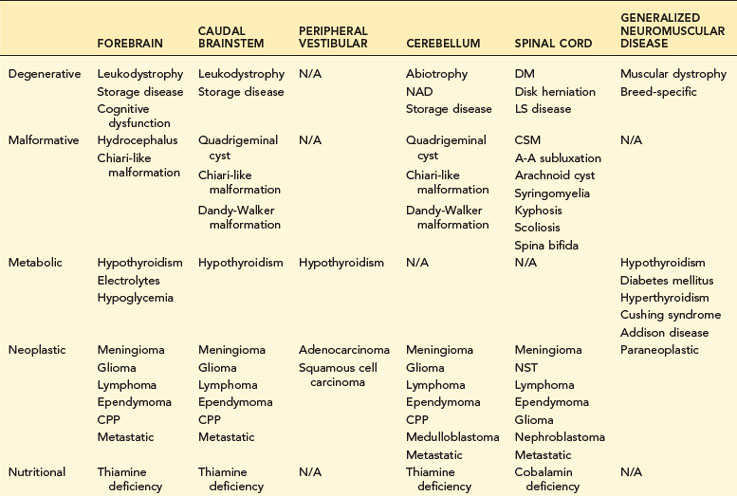
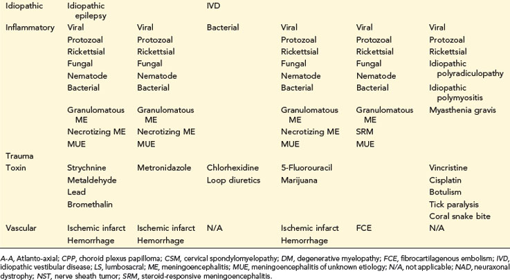
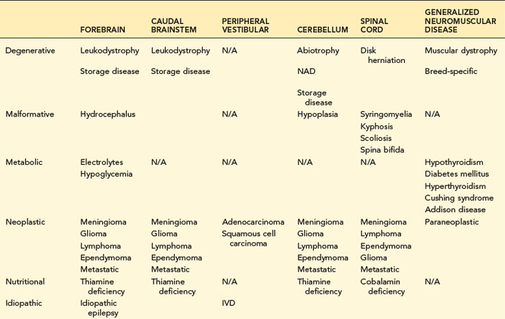
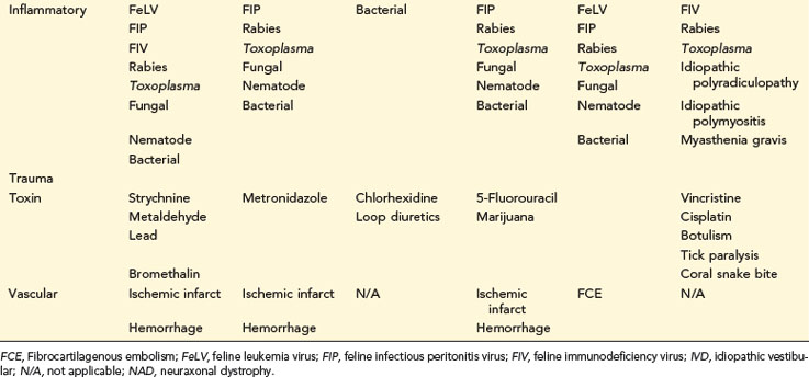
Differential diagnosis lists are based on neuroanatomic localization, signalment, as well as onset, progression, and duration of clinical signs (Tables 14-1 and 14-2). Neoplastic diseases, disk herniation, and idiopathic epilepsy all have age and breed predilections. Vascular diseases of the CNS usually have a rapid onset and are nonprogressive, whereas CNS degenerative diseases are insidious in their onset and clinical course. Appropriate neuroanatomic localization coupled with a strong differential list will allow clinicians to formulate a diagnostic plan.


Neuroimaging


Electrodiagnostics


“Electrodiagnostics” refers to a group of techniques that measure spontaneous and evoked activity arising from muscles, nerves, and CNS structures. Electromyography (EMG), motor nerve conduction (MNC), and sensory nerve conduction (SNC) are commonly performed studies to evaluate the peripheral nervous system (PNS). Somatosensory evoked potentials (SSEPs), motor evoked potentials (MEPs), electroencephalograms (EEGs), and brainstem auditory evoked responses (BAERs) are used to evaluate the CNS. In modern veterinary practices, EEG is typically used to aid in seizure detection and as a means to qualify cerebrocortical activity in animals suspected of brain death. BAER can determine integrity of auditory pathways in animals suspected of deafness or severe caudal brainstem injury.


Electrodiagnostics (i.e., EMG, MNC, SNC) are critical in the evaluation of animals with PNS disease. Presence of abnormal spontaneous activity on EMG is usually associated with axonal or muscle pathology, as opposed to junctional diseases such as myasthenia gravis. Slowing of MNC velocity may suggest a myelin disorder or loss of large-diameter axons, whereas reduction in compound motor unit action potential amplitude may indicate myopathy, axonopathy, or junctional disease. By combining results from EMG, MNC, sensory studies, repetitive nerve stimulation, and other studies, a clinician trained in electrophysiology can identify which regions of the PNS may have abnormalities. Electrophysiology cannot definitively diagnose etiology; further diagnostics such as serology and biopsy are required.


Cerebrospinal Fluid Analysis


Common Indications


Cerebrospinal fluid (CSF) is one of the only means to assess cellular responses to nervous system disease. Routine collection is suggested in animals with CNS signs or radiculopathy. In animals with intracranial CNS disease, collection is usually performed after neuroimaging (see “Contraindications”). CSF should always be acquired prior to myelography to prevent delivery of intrathecal iodinated contrast in animals with primary CNS inflammation and to avoid post-myelographic pleocytosis from hindering CSF interpretation.


Procedure for CSF Collection


CSF can be acquired from cerebellomedullary cistern (CMC) or lumbar cistern (LC) under general anesthesia. Collection from the CMC is performed by placing the animal in lateral recumbency (the side of recumbency should match the handedness of the clinician performing the procedure) on a level, secure table. The animal’s head and neck are brought to the edge of the table, and the neck is flexed approximately 90 degrees. Holding the head parallel to the tabletop is crucial for proper insertion of the collection needle and recognition of landmarks. The most commonly used technique for identifying the site of needle insertion relies on identifying the intersection of two lines, which we will call “x” and “y”. The “x” line runs from the occipital protuberance to the spinous process of the axis and defines the horizon. The “y” line runs between the cranial edges of the right and left wings of the atlas. Prior to needle insertion, the area surrounding the CSF acquisition site is clipped and sterilely prepped. A 22-gauge spinal needle (1.5 inches for dogs < 25 kg or cats; 2.5 inches for dogs > 25 kg) is then inserted through soft tissues underlying the CSF acquisition landmark. Traditionally, the bevel of the needle is directed cranially, as this may increase CSF flow rate. In small dogs or cats, the skin may need to be penetrated discretely from underlying soft tissues. The needle is advanced parallel to the table at an angle to match the mandible (often slightly caudally directed). Typically a decrease in resistance is felt after the needle penetrates the interarcuate ligament and dura, which indicates the CMC has been entered. This decrease in resistance is not always appreciated, and frequent removal of the needle stylet or advancing the needle without the stylet to check for CSF can be helpful.


Collection of CSF at the LC requires positioning the dog in lateral recumbency and flexing the lumbar vertebral column by flexing the pelvic limbs toward the ventral abdomen. The cranial wings of the ilium are located at the level of L6. Typically, a 22-gauge spinal needle (2.5 to 6 inches, dependent on dog size) is inserted parallel to the L6 spinous process to enter the L5-L6 interarcuate space. The needle is usually inserted until it contacts the ventral floor of the vertebral canal.


Most clinicians prefer CMC to LC collection, as it is easier to perform, is less frequently contaminated with blood, allows for larger volumes of CSF to be obtained, and is often easier to interpret in the setting of CNS disease because data concerning expected characteristics are more abundant. Theoretically LC CSF may be preferable for focal disease caudal to the CMC.

Stay updated, free articles. Join our Telegram channel

Sep 10, 2016 | Posted by in SMALL ANIMAL | Comments Off on Neurologic Disorders

Full access? Get Clinical Tree

Get Clinical Tree app for offline access