CHAPTER | 4 Fungal Skin Diseases


Malasseziasis (Malassezia dermatitis)
Diagnosis
Treatment and Prognosis
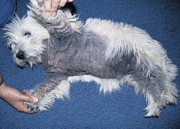
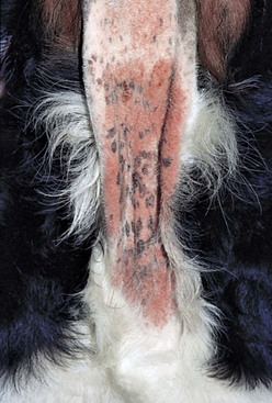
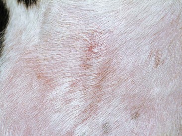
FIGURE 4-2 Malasseziasis.
Alopecia, erythema, and lichenification on the ventral neck of an allergic dog.
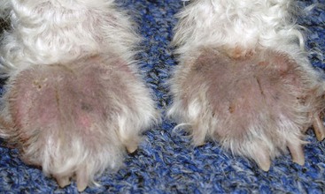
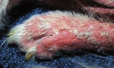
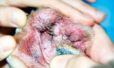
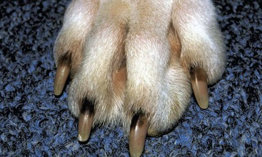
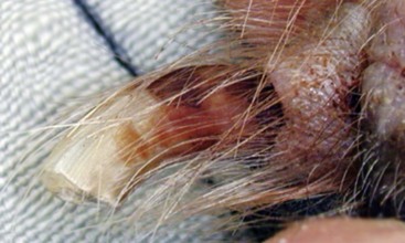
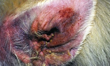
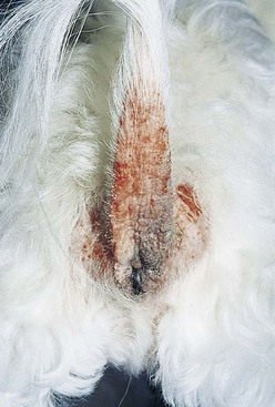
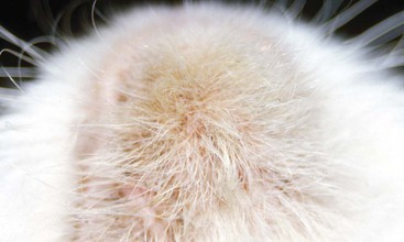
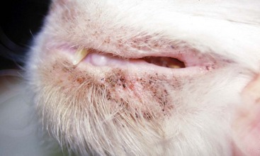
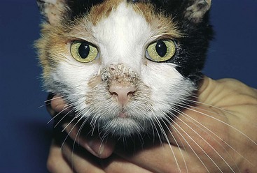
FIGURE 4-11 Malasseziasis.
The perioral dermatitis in this cat was caused by a secondary Malassezia infection.
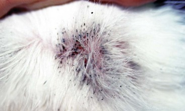
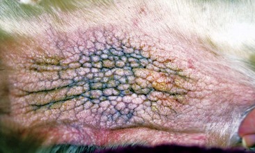
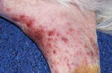
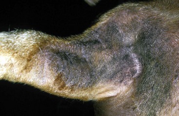
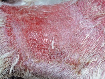
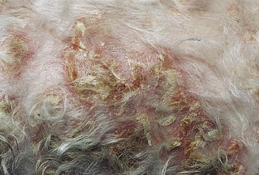
FIGURE 4-15 Malasseziasis.
More typical yeast dermatitis of the forearm compared with Figure 4-14. The alopecia, hyperpigmentation, and lichenification (“elephant skin”) are highly characteristic of yeast dermatitis.

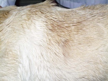
FIGURE 4-17 Malasseziasis.
Close-up of the dog in Figure 4-16. Brown discoloration of the feet is apparent and represents an early change caused by the Malassezia infection.
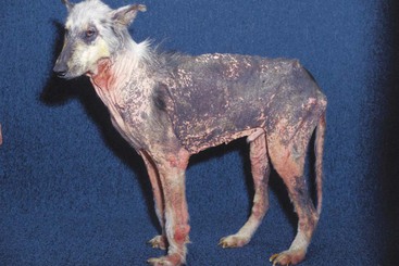
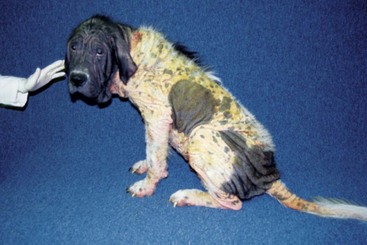
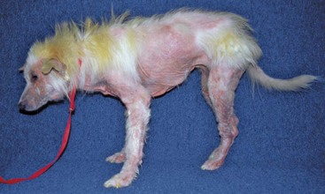
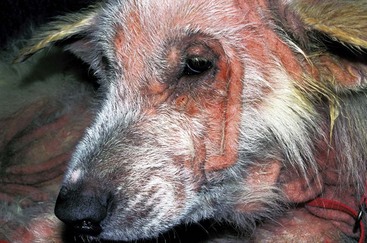
FIGURE 4-22 Malasseziasis.
Close-up of the dog in Figure 4-20. Intense erythema and alopecia, with early lichenification caused by the hypersensitivity reaction to the yeast, are apparent.
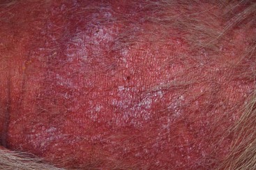
FIGURE 4-23 Malasseziasis.
Close-up of the dog in Figure 4-20. Intense erythema and alopecia caused by the hypersensitivity reaction to yeast can be seen on the thorax. Note: The skin is beginning to become lichenified, typical of Malassezia dermatitis.
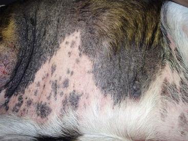
FIGURE 4-24 Malasseziasis.
Severe alopecia and lichenification typical of yeast dermatitis on the ventrum of an allergic dog.
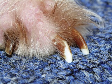
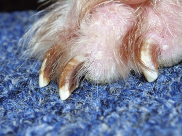
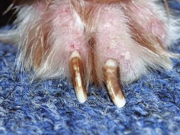
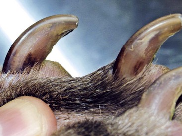
FIGURE 4-27 Malasseziasis.
Brown splotchy discoloration of the nails is a unique symptom of yeast dermatitis.

FIGURE 4-29 Malasseziasis.
Severe erythema and alopecia in the axilla of a dog. Note the moist exudate starting to form.
Candidiasis (candidosis, thrush)
Diagnosis
Treatment and Prognosis


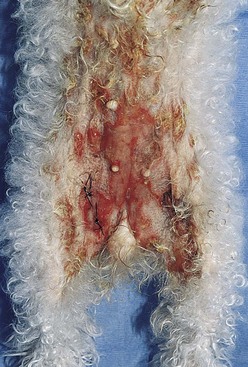
FIGURE 4-34 Candidiasis.
Superficial moist, erosive lesions on the ventrum of the dog.
(Courtesy of A. Yu.)
Dermatophytosis (ringworm)
Features
Skin involvement may be localized, multifocal, or generalized. Pruritus, if present, is usually minimal to mild but occasionally may be intense. Lesions usually include areas of circular, irregular, or diffuse alopecia with variable scaling. Remaining hairs may appear stubbled or broken off. Other symptoms in dogs and cats include erythema, papules, crusts, seborrhea, and paronychia or onychodystrophy of one or more digits. Rarely, cats present with miliary dermatitis or dermal nodules (see “Dermatophytic Granulomas and Pseudomycetomas”). Other cutaneous manifestations in dogs include facial folliculitis and furunculosis resembling nasal pyoderma, kerions (acutely developing, alopecic, and exudative nodules) on the limb or face, and truncal dermal nodules (see “Dermatophytic Granulomas and Pseudomycetomas”). Asymptomatic carrier states (subclinical infection) are common in cats, especially among long-haired breeds. Asymptomatic disease, although rare in dogs, has been reported in Yorkshire terriers.
Diagnosis
Treatment and Prognosis


Effective systemic antifungal drugs include the following:


 Itraconazole (Sporanox) 5–10 mg/kg PO q 24 hours with food
Less effective systemic antifungal drugs include the following:


Microsporum canis is one of the most common zoonotic diseases in veterinary medicine.
Adopted kittens should be screened for infection during the first veterinary wellness visit.
Even long-standing and severe infections can be resolved with aggressive and persistent treatment.
Discontinuation of therapy MUST be based on negative cultures.
Stay updated, free articles. Join our Telegram channel

Full access? Get Clinical Tree


