Diagnostic Techniques

CHAPTER | 2 Diagnostic Techniques


Diagnostic Testing


The dermatologic diagnostic minimum database includes skin scrapes, otic swabs, and cutaneous cytology. The goal should be to identify all secondary infections (e.g., pyoderma, demodicosis, dermatophytosis, otitis, Malassezia dermatitis, infectious pododermatitis), then formulate a diagnostic plan for identifying and controlling the underlying/primary disease (i.e., allergies, endocrinopathies, keratinization defects, and autoimmune skin diseases) (Box 2-1).
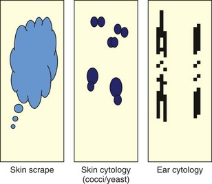
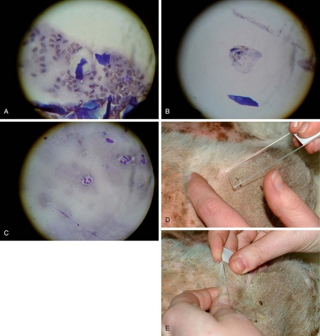
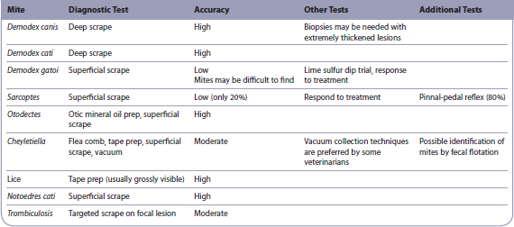
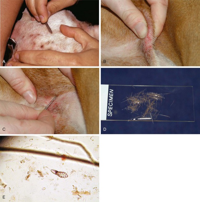


Skin Scrapes


Skin scrapes are the most common dermatologic diagnostic tests (slide #1 in the 3-slide technique). These relatively simple and quick tests can be used to identify many types of parasitic infections (Table 2-1). Although they are not always diagnostic, their relative ease and low cost make them essential tests in a dermatologic diagnostic minimum database.


Many practitioners reuse scalpel blades when performing skin scrapes; however, this practice should be stopped because of increased awareness of transmittable diseases (e.g., Bartonella, Rickettsia, feline leukemia virus [FeLV], feline immunodeficiency virus [FIV], herpes, papillomavirus).


Procedure


Deep Skin Scrapes (for Demodex spp. except D. gatoi)


A dulled scalpel blade is held perpendicular to the skin and is used with moderate pressure to scrape in the direction of hair growth. If the area is covered with hair (usually, alopecic areas caused by folliculitis are selected), it may be necessary to clip a small window to access the skin. After several scrapes, the skin should appear pink, with the capillaries becoming visible and oozing blood. This ensures that the material collected comes from deep enough within the skin to allow the collection of follicular Demodex mites. Most people also squeeze (pinch) the skin to express mites from deep within the follicles into a more superficial area, so that they may be collected more easily. If scraping fails to provide a small amount of blood, then the mites may have been left in the follicle, resulting in a false-negative finding. In some situations (with Shar peis or deep inflammation with scarring), it may be impossible to scrape deeply enough to harvest Demodex mites. These cases are few in number but require biopsy for identification of mites within the hair follicles. Hair-plucks from an area of lesional skin may be used to help find mites, but the accuracy of this technique compared with skin scrapes is unknown.


Regardless of the collection technique used, the entire slide should be searched for mites with the use of low power (usually a 10× objective). A search of the entire slide ensures that if only one or two mites are present (as is typical of scabies infection), the user will likely find them. It may be helpful to lower the microscope condenser; this provides greater contrast to the mites, thereby enhancing their visibility. (One must be sure to raise the condenser before looking for cells or bacteria on stained slides.)


Author’s Note


Cutaneous Cytology


Cutaneous cytology is the second most frequently employed dermatologic diagnostic technique (slide #2 in the 3-slide technique). Its purpose is to help the practitioner to identify bacterial or fungal organisms (yeast) and assess the infiltrating cell types, neoplastic cells, or acantholytic cells (typical of pemphigus complex).


Procedure


Fine Needle Aspirate Method


A needle (22–25 gauge) and a 6-mL syringe should be used to aspirate the mass. The area should be cleaned if necessary with alcohol or chlorhexidine. The lesion is then immobilized. The practitioner should insert the needle into the nodule while aiming for the center of the lesion, pull back on the plunger to apply suction, release and redirect, pull back on the plunger again, and stop if any blood is visible in the hub of the needle, as this will dilute the cellular sample. Negative pressure should be released before the needle is removed from the lesion. An alternative technique involves repeated insertion of the needle without the syringe into the lesion, while redirecting several times. This latter technique (without negative pressure), which decreases the frequency of inadvertent dilution of the sample with blood, works best for soft masses. Once the sample has been collected, the material is expressed onto a microscope slide by blowing a syringe-full of air through the needle to spray the cells onto the slide. The material is smeared gently to thin the clumps of cells stained with cytology stain. The slide should be scanned with low power (4×–10×) to reveal a suitable area for closer examination. A high-power (40×) objective may be used to reveal the infiltrating cell type and the cellular atypia.


Author’s Note


Acetate Tape Preparations


Tape preps are used to evaluate a variety of different conditions. The basic technique involves the use of crystal clear tape (single- or double-sided) to collect a sample of hair or superficial skin debris.


Tape Preps for Yeast


Tape preps for yeast dermatitis are some of the most efficient and effective methods of identifying Malassezia skin infections. Although they are not as reliable and quantitative as impression cultures that use Sabouraud’s media, the speed and ease of tape preps for yeast make them the techniques most commonly employed for identifying Malassezia. The lichenified lesion (elephant skin on the ventral neck or ventrum) is sampled by repeated application of the sticky side of the tape onto the lesion. The tape is then adhered to a glass slide and is stained with a cytology stain (while the first alcohol stain solution is admitted). The tape serves as a coverslip and can be examined under high power (100× oil immersion) for visualization of Malassezia organisms. This technique is useful, but false-negative results are common with all yeast collection techniques.


Stay updated, free articles. Join our Telegram channel

Sep 10, 2016 | Posted by in SMALL ANIMAL | Comments Off on Diagnostic Techniques

Full access? Get Clinical Tree

Get Clinical Tree app for offline access