47 Fundus – introduction
The fundus is the part of the posterior segment of the eye which is examined with an ophthalmoscope. It consists of the retina and choroid together with the optic disc (optic nerve head). In some animals, especially those with little pigment (e.g. merle collies), the sclera can also be seen.
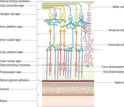
The retina is a very complicated structure and consists of 10 layers, illustrated in Figure 47.1. Nine of these layers make up the neurosensory retina while the 10th and outermost layer is the retinal pigment epithelium. More than one layer is usually involved in any pathological process but involvement of the photoreceptor layer, i.e. the rods and cones, is frequently what results in impaired vision or even blindness.
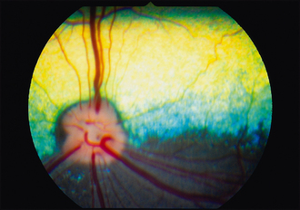
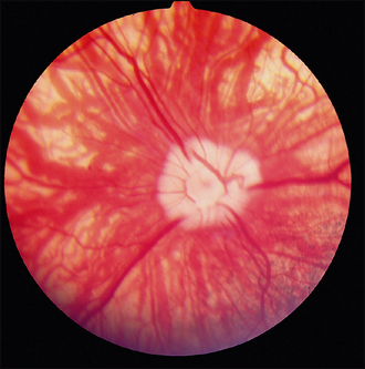
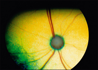
The appearance of the normal fundus varies tremendously in dogs, less so in cats (see Figures 47.2–47.5). In most animals the tapetum is present in the dorsal half of the fundus (the reflective layer within the choroid underlying the retina). The non-tapetal fundus makes up the rest of the area and is usually pigmented, due to the presence of melanin within the retinal pigment epithelium which is the outermost layer of the retina. Thus, different colours to the tapetum, absence of the tapetum, different degrees of pigmentation in the non-tapetal fundus, various shapes to the optic disc and a range of vascular patterns are all encountered. The optic disc is usually myelinated in dogs, resulting in a whitish fluffy appearance, whereas in cats it is smaller and darker due to a normal lack of myelin.
Stay updated, free articles. Join our Telegram channel

Full access? Get Clinical Tree


