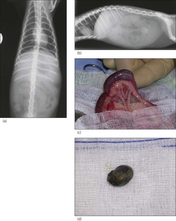
5 Ferret with abdominal mass
Introduction
This case embodies a frequent presentation in ferrets of weight loss, along with the common finding of an abdominal mass. The aim is to discuss possible investigations and outline treatment options for common conditions. (See Box 5.1 for a brief guide to ferrets in various parts of the world.)
BOX 5.1 Ferrets around the world
• In the UK, ferrets are kept for two reasons. Some are purely pet animals, some are used for working (‘ferreting’ utilizes the animals to hunt wild rabbits), and some are a combination of the two.
• Across the rest of Europe and in Russia, ferrets are variously kept as pets, for fur and for hunting.
• Although banned in certain states in the USA, in others, ferrets are extremely popular as pets. They have also been used for medical research and the fur trade for many years.
Case History
The companion ferret and other pets in the household did not show any signs of illness.
Clinical Examination
A clinical examination was performed, with the following findings:
• No abnormalities were detected on thoracic auscultation. The heart rate was 360 beats per minute (normal range in an adult ferret is 180–250)
DIFFERENTIAL DIAGNOSES
The differentials for an abdominal mass (often with weight loss) in a ferret are:
• Neoplasia (often occurs secondary to inflammatory disease):
• Adrenal gland, e.g. adrenocortical adenoma, adenocarcinoma, teratoma. This case did not show any of the usual signs seen with functional adrenal gland tumours (e.g. alopecia, pruritus or behavioural changes). Adrenal enlargement may be non-neoplastic, e.g. hyperplasia
• Pancreatic, adenocarcinoma, insulinoma (although insulinomas are often small and the clinical signs of weakness and seizures do not correlate with this case)
• Aleutian disease (viral): mass could be due to splenomegaly, mesenteric lymphadenopathy, hepatomegaly; disease also results in chronic wasting, diarrhoea, lethargy, anorexia and polydipsia
• Thickened intestines may be palpated, e.g. intestinal lymphoma, eosinophilic granulomatous disease (uncommon), proliferative bowel disease (rare, caused by Lawsonia intracellularis, mostly animals <1.5 years old)
• Several conditions will result in mesenteric lymphadenopathy that may be palpable, e.g. intestinal lymphoma, inflammatory bowel disease, eosinophilic granulomatous disease (eosinophilic infiltration and granulomas of the abdominal lymphatics and organs), proliferative bowel disease, systemic infection, tuberculosis
• Renomegaly: renal cysts, acute renal failure, hydronephrosis (e.g. associated with urolithiasis obstructing kidney, ureteral or urethral blockage)
• Mycotic disease (usually causes lung pathology, but may affect abdominal organs) – blastomycosis (splenomegaly), coccidiomycosis (various organs), histoplasmosis (splenomegaly)
• Prostatic disease (hobs), e.g. prostatomegaly with secondary urethral obstruction, prostatic abscess (may be in association with transitional cell tumours of the bladder)
BOX 5.2 Lymphoma in ferrets
• Often arises in the mesenteric lymph nodes, but may arise in gastrointestinal mucosa or metastasize there from elsewhere.
• Several types: lymphosarcoma (solid tissue tumours in organs or lymph nodes), lymphocytic leukaemia (neoplastic cells in both bone marrow and peripheral blood, acute or chronic forms)


Stay updated, free articles. Join our Telegram channel

Full access? Get Clinical Tree


