Feline uveitis

34 Feline uveitis


CLINICAL EXAMINATION


A general clinical examination should check for evidence of trauma, particularly if only one eye is affected. If both eyes are involved then systemic disease is more likely and a detailed physical examination is required. Ophthalmic examination will follow the standard format. Most cats are visual. Light reflexes are often sluggish and pupils can be fixed due to secondary synechia formation. As such, a miotic pupil, considered typical in acute uveitis, might not be present.


Some conjunctival hyperaemia and/or episcleral congestion will be present but the outside layers of the eye are not as dramatically red as in the case of canine uveitis. Instead the redness is normally inside the eye. The iris will be inflamed; rubeosis iridis is engorgement of the iridal blood vessels and neovascularization of the iris. This is noticeable as redness in a gold iris, but might manifest as a change in iris colour – from blue to green for example in cats with blue irides. Hyphaema might also make the eye appear red, but again this is less common in chronic cases.


The eye will appear cloudy. This can be due to a combination of aqueous flare, keratic precipitates, hypopyon, cataract and vitreal opacification (Figure 34.1). Oblique examination of the eye, but with the light source directed perpendicular, will assist in locating the site of opacity. Keratic precipitates tend to accumulate ventrally on the corneal endothelium. When the cat is on the examination table and you are looking down at it these opacities might not be obvious. However, if you get the cat to sit at the edge of the table and look down, while you squat on the floor and look up into its eyes, the ventral half of the eye will be more easily examined. Also when looking for keratic precipitates it is important to check both eyes. Many cases of uveitis present unilaterally but on careful examination it is clear that both eyes are affected to differing degrees.


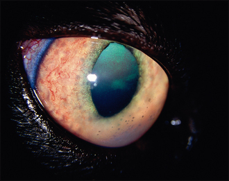
Aqueous flare will vary from just a slight haze to the anterior chamber through to full blown hypopyon obscuring further intraocular examination. Similarly, most cats will have a clear lens, but in chronic cases a total mature cataract can be present. Vitreal inflammation, hyalitis, is quite common in chronic uveitis cases and will most frequently be centred behind the lens – a pars planitis. Without careful examination this can be confused with cataract. Some cases of anterior uveitis will also have chorioretinitis as well. Thorough fundus examination, after pupil dilation with tropicamide, might reveal both active and inactive chorioretinal lesions.


The cornea should be routinely stained with fluorescein – traumatic incidents can result in ulceration, plus it is important to ensure that no active corneal damage is present since this could alter the treatment regime. Intraocular pressures should also be checked if possible. Uveitis results in a lowered intraocular pressure but chronic cases in cats frequently develop secondary glaucoma which might not be obvious on the ophthalmic examination alone.


CASE WORK-UP


If the case appears traumatic, minimal further work-up is required. However, most cases do not present with an obvious cause and, as such, diagnostic tests are normally required. The clinical appearance is the same regardless of cause, and thus one cannot determine which virus is involved, for example, on ophthalmic appearance alone. A baseline investigation would therefore include full haematology and biochemistry (looking for evidence of infection, renal disease, chronic anaemia for example) and viral screening.


FeLV and FIV status should always be checked. Toxoplasma titres (both IgM and IgG) are important in cats which hunt, fight or show any retinal changes. If the cat is FeLV positive then it should be carefully evaluated for evidence of systemic lymphoma – abdominal ultrasound should be performed for example. FIP titres and a coronavirus screen should be considered in young animals and cats which are systemically ill, but if only ocular changes are present in adults then this is a less likely cause for the uveitis.


Other infectious agents such as Bartonella henselae can be considered if the preliminary screening does not provide any definitive answers. Thankfully systemic fungal infections in cats in the UK are very rare, but can be considered if the cat has travelled from endemic countries. Unfortunately, despite extensive diagnostic testing, many cases remain idiopathic. On occasion it is useful to perform aqueocentesis to try to establish a definitive cause but such cases should be referred to a suitably qualified specialist – the risks of further iatrogenic damage are high even in experienced hands.


Sep 10, 2016 | Posted by in SMALL ANIMAL | Comments Off on Feline uveitis

Full access? Get Clinical Tree

Get Clinical Tree app for offline access