Feline corneal sequestrum

30 Feline corneal sequestrum


CLINICAL EXAMINATION


General clinical examination is usually unremarkable, unless an active FHV-1 infection is concurrent. Ophthalmic examination will reveal several abnormalities. The affected eye is usually wet, and Schirmer tear test readings will confirm this with a higher reading than in the normal eye. However, on occasion the opposite can be true – a sticky discharge might suggest an associated keratoconjunctivitis sicca so tear test readings should not be overlooked.


Blepharospasm is normally present. Conjunctival hyperaemia and some chemosis are common, as is superficial corneal vascularization. A darkly pigmented patch of cornea defines the sequestrum. This can be raised above the epithelium or subepithelium (the latter is less uncomfortable). The colour can vary from a mid brown to almost black. The ocular discharge is often also darkly stained; however, this is normal for many cats so should not be over-interpreted. Corneal ulceration is often superficial or mid-stromal and fluorescein dye should always be used for any suspected sequestrum. Variable amounts of corneal vascularization can be encountered – in some patients none is noted, while in others there is profuse superficial vascularization and even granulation tissue deposits at the edge of the sequestrum.


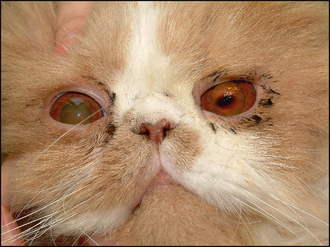
In more severe cases the pupil can be miotic, with rubeosis iridis and some aqueous flare – a reflex uveitis – which is most common if bacterial infection is present with the surface disease. In long-standing cases there might be some degree of secondary lateral lower lid entropion with the hairs rubbing on the cornea and exacerbating the problem – this is more frequent in older domestic short haired breeds and is rarely seen in Persians. However, in this latter breed a medial lower lid entropion can be present which may contribute to the problem. In addition, brachycephalic cats can suffer from a relative lagophthalmos such that the central cornea is not fully protected during blinking and tear film dynamics are abnormal (Figure 30.1). This is a serious problem in some of the ‘ultra-type’ Persians with the excessively flat face and bulging eyes – some of these poor cats even sleep with their eyes partially open and corneal health can be severely compromised as a result.


Checking the frequency of blinks, and whether these are complete or partial – where the eyelids do not fully meet – is important. In some patients, particularly the predisposed breeds, both eyes can be affected although often not at the same time, and certainly rarely symmetrically if both are involved.


Sep 10, 2016 | Posted by in SMALL ANIMAL | Comments Off on Feline corneal sequestrum

Full access? Get Clinical Tree

Get Clinical Tree app for offline access