53 Exophthalmos
CLINICAL EXAMINATION
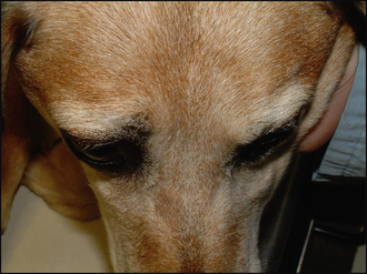
On ophthalmic examination the animal will usually have an obviously abnormal appearance. The face will be asymmetrical in unilateral cases. The affected globe might appear enlarged, but on close examination it will be seen to be pushed forwards. Thus, looking at the patient both from above and the side, as well as from in front, will assist in determining the position of the globe (Figure 53.1). The globe might be deviated in position (squint or strabismus) and this is most commonly in a lateral or dorsolateral direction (the deviation will assist in determining the location of any retrobulbar mass). The third eyelid is normally elevated and some ocular discharge is frequent. This can vary from epiphora to a frank purulent discharge. Sometimes it can be bloodstained. Resistance to retropulsion will be present, and this might be painful in some animals. The globe should be gently pushed backwards behind the closed lids and the two sides compared. The amount of normal retropulsion possible varies from breed to breed – it is quite extensive in breeds such as the Great Dane and Rottweiler, while it is minimal in brachycephalic dogs and cats.
Any animal with exophthalmos should have a thorough oral examination. This will concentrate on evidence of dental disease, including gingivitis, examination of the back of the mouth behind the last molar (looking for swelling, hyperaemia, discharge and abnormality of the oral mucosa), and checking for wounds or retained foreign bodies. Many patients will exhibit pain when opening the mouth (as the vertical ramus of the mandible impinges on the retrobulbar space). A nasal discharge might be present – for example, if a caudal nasal tumour is causing the exophthalmos. Alternatively, nasal air flow might be reduced and comparison of the two sides can be useful – holding a small tuft of hair in front of each nostril in turn to visualize the air flow on expiration can be helpful.
Table 53.1 provides differential diagnoses for the three most common causes of exophthalmos.
Table 53.1 Differential diagnosis of the common causes of exophthalmos
| Abscess or cellulitis | Neoplasia | Myositis |
|---|---|---|
CASE WORK-UP
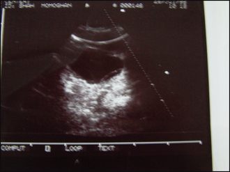
Orbital ultrasonography can be rewarding, and usually can be performed in the conscious patient with topical anaesthesia only, although some patients might require sedation. A 7.5–10 MHz sector transducer is coated with normal viscous ultrasound gel and is placed directly on the desensitized cornea (no stand-off is required). The retrobulbar space will be demonstrated. Imaging through the lids will result in a less clear image as trapped air between the eyelid hairs will cause interference. A normal retrobulbar space will consist of a cone shape of hyperechoic (bright) fat and hypoechoic (dark) muscle extending to the apex of the orbit. Sometimes a dark central line can be demonstrated which corresponds to the optic nerve. Medially the hyperechoic curving structure represents the orbital bone. The retrobulbar space should be examined in both horizontal and vertical planes, with comparison to the fellow eye being useful if one is uncertain of the normal appearance. A retrobulbar mass will appear as a region of hypo- or mixed echogenicity within this cone.
Unfortunately, ultrasonography cannot indicate histological type. However, a solid hyperechogenic mass is more likely to be neoplastic than a mixed echo – where the heterogeneous echo represents both solid and liquid such as is seen with an abscess. If the back of the globe is indented by a mass, this is more common with neoplastic processes than inflammatory ones (Figure 53.2). Occasionally a foreign body can be identified by virtue of the acoustic shadow it casts. Swollen extraocular muscles can be highlighted by ultrasonography in cases of extraocular muscle myositis. Ultrasound-assisted fine needle aspiration can be performed, but this can be dangerous (resulting in inadvertent globe rupture for example) and is best left to specialists.
Stay updated, free articles. Join our Telegram channel

Full access? Get Clinical Tree


