Esophagoscopy

Chapter 3 Esophagoscopy


The term esophagoscopy refers to examination of the lumen and mucosal lining of the esophagus with endoscopic equipment. In most cases, esophageal disease is diagnosed by the clinical history combined with contrast radiography of the esophagus, esophagoscopy, or both. Radiography is the preferred method for documenting megaesophagus, motility dysfunction, vascular ring anomalies, and hiatal hernia. Esophagoscopy is most reliable for the diagnosis of disorders that disrupt the mucosa or obstruct the lumen, such as esophagitis, stricture, foreign body, and neoplasia. Esophagoscopy not only is a valuable diagnostic procedure but also is useful as a therapeutic intervention for removing foreign bodies, performing balloon or bougie dilation of strictures, placing indwelling feeding tubes, placing endoluminal stents, and assisting laser treatment of esophageal tumors.


Indications


Esophagoscopy is indicated for the diagnostic evaluation of animals with signs of esophageal disease, including regurgitation, dysphagia, odynophagia, and excessive salivation. Esophagoscopy is also indicated for evaluation of animals known or suspected to have ingested a potential foreign body (see Chapter 7). This minimally invasive procedure allows visual examination of the esophageal mucosa and lumen and facilitates the procurement of specimens for biopsy, cytology, and culture. Thus esophagoscopy is most effective for obtaining a definitive diagnosis of conditions involving the mucosa or abnormalities within the lumen, including esophageal foreign body, esophagitis, esophageal stricture, esophageal neoplasia, and gastroesophageal intussusception. Compared with contrast radiography, esophagoscopy is less definitive for diagnosing megaesophagus and other motility disorders, diverticulum, hiatal hernia, and compression by periesophageal masses, although it often provides valuable diagnostic information in these situations. Esophagoscopy can also be used as therapeutic intervention to guide balloon catheters for dilating esophageal strictures, to assist deployment of esophageal stents, to remove esophageal foreign bodies (see Chapter 7), to place indwelling gastrostomy (see Chapter 9) or esophagostomy feeding tubes, and to ablate neoplastic tissue with lasers.


Esophagoscopy is most often indicated for diagnostic evaluation of patients with regurgitation. Regurgitation is the passive retrograde expulsion of food or fluid from the esophagus. Regurgitation must be distinguished from vomiting, which is a centrally mediated reflex characterized by the forceful ejection of gastroduodenal contents preceded by hypersalivation, retching, and abdominal contractions. The timing of regurgitation in relation to eating is determined by the location of the esophageal abnormality, the presence and degree of obstruction, and the reservoir effect of esophageal dilation. Regurgitation immediately after eating is most likely to occur with proximal esophageal lesions or complete esophageal obstruction. However, regurgitation may be unassociated with eating when dilation of the esophagus provides a reservoir for prolonged retention of food and fluid. The selective retention of fluids over solid food is more likely to occur with partial obstruction. Regurgitated material is usually composed of undigested food (often tubular) and white to clear frothy liquid (mucus and saliva), but fresh blood may be seen with mucosal trauma or erosions. Putrefaction of food may occur after prolonged sequestration in a dilated esophagus. In comparison, vomitus usually consists of partially digested food mixed with bile-stained fluid. Both regurgitation and vomiting may be observed in animals with hiatal hernia, gastroesophageal intussusception, and acid-induced esophagitis secondary to severe vomiting. Hematemesis is usually a sign of gastric disease but is occasionally seen in animals with esophageal neoplasms that are bleeding extensively.


The age of onset is important because regurgitation beginning at the time of weaning suggests a vascular ring anomaly, congenital idiopathic megaesophagus, or congenital stenosis. An acute onset of regurgitation suggests the presence of an esophageal foreign body or acute esophagitis, whereas a chronic history of regurgitation is more consistent with idiopathic megaesophagus, vascular ring anomaly, hiatal hernia, chronic reflux esophagitis, or esophageal neoplasia. Intermittent signs are often seen in animals with hiatal hernia and reflux esophagitis. The history may indicate potential exposure to foreign bodies or caustic medications (e.g., doxycycline or clindamycin), a recent anesthetic procedure that could cause reflux esophagitis and esophageal stricture, or signs of neurologic or neuromuscular dysfunction that could be associated with secondary megaesophagus.


Dysphagia, or difficulty in swallowing, is another indication for esophagoscopy. Difficulty in swallowing often suggests the presence of oropharyngeal disease, cranial esophageal obstruction, a motility disturbance, or pain caused by a foreign body, stricture, esophagitis, or another abnormality in the cranial esophagus. Dysphagia is usually associated with repeated or exaggerated attempts to swallow, odynophagia (pain on swallowing), and ptyalism (excessive salivation). Dysphagia can be characterized by observing the animal while it is drinking and eating both dry and canned food. Unexplained salivation is also an indication for esophagoscopy. In some animals with esophagitis or neoplasia (especially early in the course), unexplained salivation may be the most apparent clinical sign. Dysphagia and ptyalism must also be differentiated from oropharyngeal and salivary gland causes, such as sialadenitis and sialadenosis.


Other clinical signs of esophageal disease can also be indications for esophagoscopy. Weight loss occurs secondary to inadequate food intake and is related to the severity of esophageal dysfunction. An otherwise healthy animal with persistent regurgitation may have a ravenous appetite because of an inability to retain ingested food. This situation is common in animals with a megaesophagus, vascular ring anomaly, or esophageal stricture. In contrast, anorexia can occur in animals that have painful and difficult swallowing associated with severe esophagitis, an esophageal foreign body, or neoplasia. Anorexia can also occur in conjunction with coughing, dyspnea, and fever in animals that have secondary aspiration pneumonia, esophageal perforation, or a bronchoesophageal fistula. Occasionally, laryngeal stridor and change or loss of voice as a result of chronic laryngitis from exposure to gastric acid may be the primary presenting signs of severe gastroesophageal reflux. Hypomotility and megaesophagus have been associated findings in dogs with laryngeal paralysis.


The clinical signs of diseases of the esophagus, stomach, and intestinal tract often overlap, and diseases may involve more than one region of the digestive tract. As a result, endoscopic examinations limited to the esophagus are not usually appropriate. Most animals that undergo esophagoscopy should have a thorough endoscopic examination of the entire upper gastrointestinal tract so that the information necessary for an accurate assessment can be obtained and so that associated or unsuspected abnormalities can be detected in other regions. This complete examination is termed esophagogastroduodenoscopy (EGD).


Instrumentation


Esophagoscopy can be performed with flexible or rigid endoscopes; however, flexible video endoscopes are most versatile because of their superior optics, illumination, insufflation, recording functions, and maneuverability. Flexible video endoscopes for examination of the gastrointestinal (GI) tract should have a working length of at least 110 cm, a diameter less than 9.8 mm, suction and air/water insufflation capability, four-way tip deflection of at least 180 degrees by 90 degrees, and a 2.0- or 2.8-mm instrument channel (see Chapter 1). Flexible endoscopes readily enable thorough evaluation of the stomach and duodenum during the same procedure, which is not possible with rigid scopes.


Rigid endoscopes for esophagoscopy are available in various lengths and diameters and are most useful for removal of certain types of esophageal foreign bodies. Even when a flexible endoscope is the primary instrument, one or two multipurpose rigid scopes should be available for selected situations, such as esophageal foreign body removal with rigid grasping instruments or flexible endoscope extraction of sharp foreign bodies with the rigid scope used as a protective “overtube” (see Chapter 7). Most rigid endoscopes marketed for procedures in people have a light source, an air insufflation mechanism, and a blunt-tipped obturator that fits inside the scope to facilitate insertion into the lumen of the esophagus. Pediatric rigid scopes have a diameter of approximately 12 mm, whereas adult rigid scopes are approximately 25 mm in diameter.


The accessory instruments needed for esophagoscopy include biopsy forceps, cytology brushes, foreign body retrieval instruments, injection needles, and a set of balloon catheters or bougies of various sizes (6- to 30-mm diameter) for dilation of esophageal strictures. Accessory instruments for foreign body removal are discussed in Chapter 7. Oval-cupped alligator jaw biopsy forceps (especially without a central spike) are adequate for obtaining mucosal biopsy specimens of the stomach, small intestine, and large intestine, but they are generally less effective for obtaining diagnostic biopsy specimens from the tough mucosa of the esophagus. Forceps with a central spike can improve the results of esophageal biopsies.


Optional accessory instruments can include equipment for electrocautery, electrosurgery, polypectomy (snares), endoluminal stenting (for strictures or inoperable tumors), and photo/laser/argon therapy for the ablation of neoplastic tissue as a palliative measure to relieve obstruction and stop hemorrhaging.


Patient Preparation and Restraint


Esophagoscopy requires general anesthesia, and food should be withheld for a minimum of 12 hours before the procedure. Throughout the procedure the anesthetized patient should have an endotracheal tube and mouth gag in place. The patient is positioned in left lateral recumbency.


When an esophageal perforation, foreign body, or stricture is suspected, additional precautions should be taken. Insufflation of air into a perforated esophagus can produce life-threatening tension pneumothorax. Foreign bodies, especially bones and other sharp objects, can cause esophageal perforation, as can iatrogenic tearing from balloon or bougie dilation of strictures. Therefore, before endoscopic retrieval of foreign bodies, it is advisable to review cervical and thoracic radiographs for signs of perforation such as increased periesophageal mediastinal density, pleural effusion, pneumomediastinum, or pneumothorax. Esophageal perforation is usually an indication for surgical exploration rather than endoscopy. If the radiographs reveal no evidence of perforation in the patient with an esophageal foreign body, the esophagoscopic examination can proceed. Thoracic radiography for pneumothorax from iatrogenic perforation is also indicated after foreign body removal or stricture dilation procedures that cause excessive esophageal trauma. During examination of esophageal obstructive lesions, such as strictures, excessive insufflation of air should be avoided because it can be difficult to evacuate the air through the stenotic area if the caudal esophagus and stomach become overdistended.


Barium contrast radiography should ideally be completed 24 hours before esophagoscopy so that retained contrast medium does not compromise the mucosal examination. A clear, water-soluble, nonionic iodinated contrast agent (e.g., iohexol [Omnipaque]) should be used instead of barium when esophagoscopy is likely to be performed within a few hours of contrast radiography. If esophagoscopy has to be done shortly after a barium esophagram, lavage of the esophagus and stomach with water may be necessary for the barium to be evacuated. If barium has pooled in the esophagus because of esophageal obstruction, hypomotility, or gastroesophageal reflux, a suction tube should be used to remove as much of the contrast medium as possible before anesthesia is induced so that the risk of barium aspiration is minimized during the procedure and so that mucosal visibility is maximized. Alternatively, an animal with a motility disorder may be held upright so that contrast material can flow into the stomach by gravity. The accessory channel of a flexible endoscope should not be used to suction a large volume of undiluted barium from the esophagus because the barium residue may adhere to the walls and occlude the channel. Furthermore, once the residue dries, it can be difficult to remove.


Esophagoscopy Procedure


The anesthetized and intubated patient is positioned in left lateral recumbency with a mouth speculum securely in place to protect the scope. The endotracheal tube is important for preventing the aspiration of refluxed or regurgitated material from the oropharynx during the procedure. During rigid endoscopy procedures the endotracheal tube also prevents collapse of the cervical trachea from the pressure of the rigid scope. The insertion tube of the endoscope should be prelubricated with water-soluble lubricant gel for ease of passage. Alternatively, the scope may be lubricated with oral secretions as it is passed into the oral cavity.


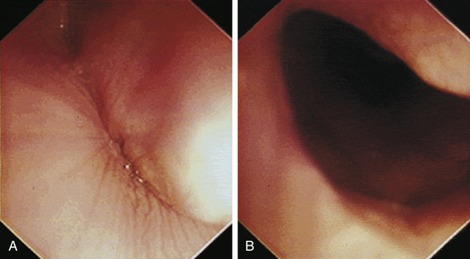
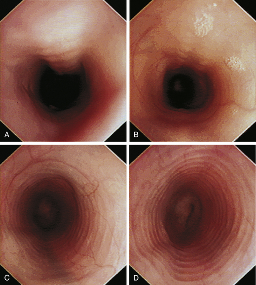
With the animal’s head and neck extended, the endoscope is directed centrally through the oropharynx and guided dorsal to the endotracheal tube and larynx so that the cranial esophageal sphincter (CES) comes into view. The CES is the entrance to the esophagus and is normally closed, appearing as a star-shaped area of folded mucosa dorsal to the larynx (see Figure 3-1, A-B). With insufflation and minimal pressure of the endoscope tip against the CES, the scope is easily advanced through the low-resistance sphincter into the cervical esophagus. Resistance is occasionally felt when the endoscope tip is misdirected into one of the piriform recesses located on either side of the larynx. Withdrawing and redirecting the endoscope more toward the dorsal midline easily corrects this problem. If the endoscope tip is passed blindly in large-breed dogs, it may inadvertently be advanced through the laryngeal opening into the proximal trachea. Resistance is felt when the endoscope comes into contact with the endotracheal tube. If this occurs, the endoscope tip should be pulled back to the oropharynx and redirected into the CES.


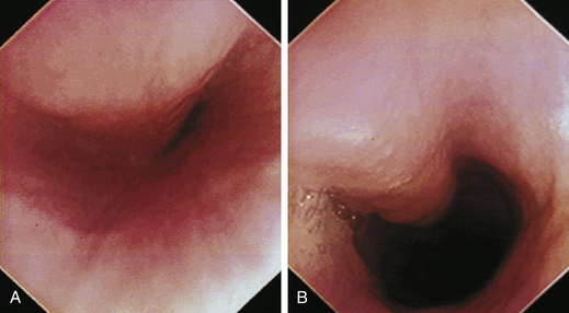
The cervical esophagus is normally collapsed, so as the endoscope passes through the sphincter, a brief “red out” usually obscures visibility. Insufflation of air should begin immediately and continue until the esophageal lumen is clearly visualized ahead of the scope as it advances (see Figure 3-2, A-B). The esophageal lumen forms a straight tube except for a slight flexure at the thoracic inlet, where the cervical and thoracic regions of the esophagus meet. The endoscope should meet little or no resistance as it is advanced. The endoscopist should advance the scope down the esophagus in a slow continuous motion, using only minor adjustments in tip deflection and torque to maintain a full panoramic view of the lumen and mucosal surfaces. Air should be insufflated intermittently, as needed, to keep the lumen distended ahead of the scope. To maintain distension in some patients, it may be necessary to have an assistant help occlude the esophagus just caudal to the larynx to prevent the escape of insufflated air. The lumen of the thoracic esophagus generally opens with minimal insufflation. Pulsations of the aorta against the wall of the esophagus are seen at the level of the base of the heart.


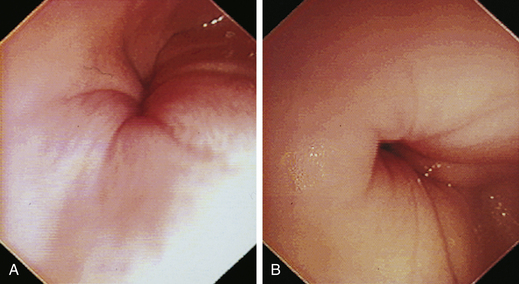
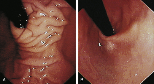
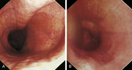
At the gastroesophageal junction the esophagus passes obliquely through the diaphragm to open into the stomach. The gastroesophageal sphincter (GES) is not a true anatomic sphincter but rather a high-pressure zone that keeps the distal esophagus closed between swallows (see Figure 3-3, A). To advance the endoscope through the slitlike opening of the GES and into the stomach, the endoscopist deflects the tip approximately 30 degrees to the left and slightly upward (see Figure 3-3, B). This can be done easily under direct visualization, and minimal or no resistance should be encountered as the endoscope advances through the GES.


A description of the normal appearance of the esophagus in dogs and cats and corresponding images are found in the “Atlas” section of this chapter (see Figures 3-1 through 3-10).


Sample Procurement


Esophageal biopsy is not usually required for diagnosis of most esophageal diseases. Visual inspection alone is generally adequate. The primary indications for mucosal biopsy of the esophagus include the presence of a mass or mucosal abnormalities indicative of esophagitis. It can be difficult to obtain adequate esophageal mucosal biopsy specimens with standard endoscopic pinch biopsy forceps, especially when the mucosa is normal. It is difficult to orient the forceps perpendicular to the wall of the esophagus, and the mucosa is often too tough to cut with the forceps, so the biopsy cups tend to slide off as they are closed. Rigid biopsy forceps with larger biopsy cups can be used with rigid endoscopy equipment to improve the quality of esophageal biopsies. Alternatively, a biopsy forceps with a central spike (bayonet type) can be used (see Figure 8-1). The spike is helpful for anchoring the biopsy forceps on the mucosa so that the “bite” is more perpendicular and slipping is prevented.


Standard biopsy forceps are most effective for obtaining adequate biopsy specimens from proliferative esophageal masses and diseased mucosa with increased friability. Six to 10 biopsy specimens are usually adequate for definitive diagnosis. Preferentially, the tissue samples should be taken from viable areas of tumors and from border areas between obvious proliferative growth and the area of invasion. Central necrotic, ulcerated areas should be avoided because the diagnostic yield is lower in these areas. Brush cytology specimens are sometimes useful for diagnosing esophageal neoplasia or characterizing unusual forms of esophagitis such as mucosal candidiasis. A guarded (sheathed) cytology brush prevents contamination and loss of specimen material as the brush is withdrawn through the accessory channel of the endoscope. Brush cytology specimens should be collected first to avoid diluting a cytology specimen with blood from a recent biopsy site.


Postoperative Care and Complications


Patients undergoing routine diagnostic esophagoscopy are recovered routinely from anesthesia and discharged the same day. Normal food and water intake can resume after a few hours once anesthetic recovery is complete. Complications related to diagnostic esophagoscopy are rare and primarily limited to inadvertent aspiration of oropharyngeal secretions or refluxed gastroesophageal fluid caused by improper protection of the airway with an endotracheal tube during the procedure and anesthetic recovery. In esophageal stricture, excessive insufflation of air should be avoided because it can sometimes be difficult to evacuate the air through the stricture if the caudal esophagus and stomach become overdistended.


Esophageal perforation is a rare but serious complication of esophageal foreign bodies (e.g., bones and other sharp objects) or their removal or the result of iatrogenic tearing of the esophagus during balloon or bougie dilation of esophageal strictures. Insufflation of air into a perforated esophagus can produce life-threatening tension pneumothorax. If the esophagus is perforated, esophagoscopy should not be performed. If esophagoscopy is under way and perforation is suspected, the procedure should be discontinued and thoracic radiographs obtained immediately. Evacuation of air or fluid from the pleural cavity may be necessary, as well as immediate surgical repair.


Most patients do not require medication after routine esophagoscopy. Postoperative medications to control pain and esophagitis are usually indicated after interventional esophagoscopy procedures such as esophageal foreign body removal and stricture dilation. Esophagitis is treated with a proton pump inhibitor for acid control, sucralfate as a mucosal protectant, and metoclopramide or cisapride as prokinetics to increase GES tone and prevent acid reflux. Analgesics are prescribed according to the anticipated level of discomfort from the procedure and associated mucosal injury.


Atlas Pages 46-94


Normal Canine Esophagus


Figure 3-1, p. 46, Cranial esophageal sphincter (closed and open)

Figure 3-2, p. 46, Cervical esophagus—partially and fully distended

Figure 3-3, p. 47, Gastroesophageal sphincter

Figure 3-4, p. 47, Midthoracic esophagus—distended

Normal Feline Esophagus


Figure 3-5, p. 48, Cervical and thoracic esophagus—distended

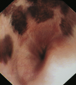
Pigmented Esophageal Mucosa in Dogs


Figure 3-6, p. 49, Pigmented esophageal mucosa in a Chow

Figure 3-7, p. 49, Pigmented esophagus in a Shar-Pei with a hiatal hernia

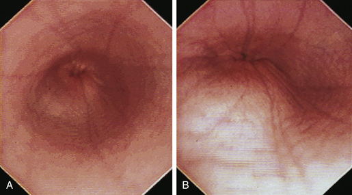
Normal Gastroesophageal Junction


Figure 3-8, p. 49, Canine caudal thoracic esophagus and closed gastroesophageal sphincter

Figure 3-9, p. 50, Feline caudal thoracic esophagus and closed gastroesophageal sphincter

Figure 3-10, p. 50, Retroflex view of gastroesophageal junction

Megaesophagus


Figure 3-11, p. 51, Congenital megaesophagus in a Great Dane—survey and contrast radiographs

Figure 3-12, p. 51, Megaesophagus in a cat—radiographs

Figure 3-13, p. 52, Megaesophagus in a dog

Figure 3-14, p. 52, Megaesophagus with marked dilation

Figure 3-15, p. 53, Megaesophagus with extensive pooling of fluid

Diverticula


Figure 3-16, p. 54, Diverticulum secondary to esophageal stricture—radiograph

Figure 3-17, p. 54, Diverticulum secondary to megaesophagus—radiograph and endoscopic view with retained ingesta and esophagitis

Figure 3-18, p. 55, Diverticulum in a cat with a trichobezoar

Vascular Ring Anomalies


Figure 3-19, p. 56, Persistent right aortic arch—schematic

Figure 3-20, p. 57, Persistent right aortic arch in a cat

Figure 3-21, p. 58, Persistent right aortic arch in a cat

Figure 3-22, p. 58, Persistent right aortic arch in a dog

Figure 3-23, p. 59, Persistent right aortic arch in a dog

Figure 3-24, p. 59, Persistent right aortic arch in a dog

Figure 3-25, p. 59, Persistent right aortic arch in an 8-year-old Labrador retriever

Esophagitis


Figure 3-26, p. 60, Severe esophagitis and hemorrhaging in a dog with a hoof-claw foreign body

Figure 3-27, p. 61, Hairball-induced esophagitis in a cat

Figure 3-28, p. 62, Reflux esophagitis in a dog with chronic vomiting

Figure 3-29, p. 62, Reflux esophagitis in a dog with chronic intermittent vomiting

Figure 3-30, p. 63, Reflux esophagitis in a dog with vascular ring anomaly

Figure 3-31, p. 63, Reflux esophagitis in a dog with hypergastrinemia and hypertrophic gastropathy

Figure 3-32, p. 63, Esophagitis after anesthesia for ovariohysterectomy

Figure 3-33, p. 63, Chemically induced erosive esophagitis

Figure 3-34, p. 64, Eosinophilic esophagitis in a cat

Figure 3-35, p. 65, Esophageal candidiasis in a dog with persistent right aortic arch

Esophageal Stricture


Figure 3-36, p. 67, Barium contrast esophagram—esophageal stricture

Figure 3-37, p. 67, Mature fibrotic esophageal stricture

Figure 3-38, p. 68, Hairball-induced esophageal stricture

Figure 3-39, p. 68, Triple esophageal stricture in a dog after general anesthesia for fracture repair

Figure 3-40, p. 69, Partial stricture with thin fibrotic ridges

Figure 3-41, p. 69, Chronic esophageal stricture with a network of fibrotic bands and sacculations

Figure 3-42, pp. 70-71, Barium contrast esophagram and esophageal stricture series—feline

Figure 3-43, p. 72, Various sizes of balloon dilators (inflated)

Figure 3-44, p. 72, Balloon inflation device with integrated pressure gauge

Figure 3-45, p. 73, Through-the-scope balloon dilator

Figure 3-46, p. 74, Videofluoroscopy sequence of stricture dilation with a guide-wired balloon

Figure 3-47, p. 75, Sequential endoscopic views of a balloon dilation procedure

Figure 3-48, p. 76, Esophageal stricture series—initial examination and balloon therapy

Figure 3-49, p. 76, Moderate to severe mucosal tearing and hemorrhaging after balloon dilation

Figure 3-50, p. 77, Rupture of an esophageal stricture in a cat

Figure 3-51, p. 78, Positioning of an endotherapy injection needle for intralesional injection

Figure 3-52, p. 79, Metal, olive-tipped bougies and Savary-Gilliard dilators

Esophageal Perforation


Figure 3-50, p. 77, Rupture of an esophageal stricture in a cat

Esophageal Fistula


Figure 3-53, p. 81, Chronic esophageal fistula after esophageal perforation by a sharp stick

Hiatal Hernia


Figure 3-54, p. 83, Severe gastroesophageal reflux in a cat with megaesophagus and hiatal hernia—contrast radiographs and endoscopic views

Figure 3-55, p. 84, Hiatal hernia in a Shar-Pei

Figure 3-56, p. 85, Hiatal hernia and reflux esophagitis—canine

Figure 3-57, p. 85, Paraesophageal hiatal hernia—canine

Figure 3-58, p. 86, Hiatal hernia and gastroesophageal intussusception—canine

Gastroesophageal Intussusception


Figure 3-59, pp. 87-88, Hiatal hernia with gastroesophageal intussusception—feline

Figure 3-60, p. 88, Gastroesophageal intussusception in a cheetah

Figure 3-61, p. 89, Gastroesophageal intussusception in a cheetah

Figure 3-62, p. 89, Gastroesophageal intussusception in a cheetah

Figure 3-63, p. 89, Gastroesophageal intussusception in a cheetah

Esophageal Neoplasia


Figure 3-64, p. 90, Squamous cell carcinoma—feline

Figure 3-65, p. 91, Papillary carcinoma—canine

Figure 3-66, p. 91, Mast cell tumor of the esophagus—canine

Figure 3-67, p. 92, Undifferentiated sarcoma—feline

Figure 3-68, p. 92, Leiomyoma at gastroesophageal junction—canine

Figure 3-69, p. 93, Mass at gastroesophageal junction—probable leiomyoma

Figure 3-70, p. 93, Lymphoma—canine

Periesophageal Masses


Figure 3-71, p. 94, Poorly differentiated sarcoma—canine


Atlas for Appearance of the Normal Esophagus


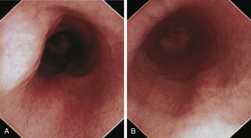
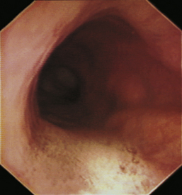
The normal esophagus in the fasted animal is empty or contains a minimal amount of clear fluid or foam (Figures 3-1 through 3-4). If the esophagus contains food residue, a large pool of fluid, or bilious fluid, then gastroesophageal reflux, hiatal hernia, motility dysfunction, or esophageal obstruction should be suspected. Because gastroduodenal contents may occasionally reflux into the esophagus during gastroduodenoscopy, the esophagus should be thoroughly assessed before advancing the scope into the stomach. In anesthetized animals the normal esophagus becomes flaccid and dilated, which makes the tubular lumen appear large when insufflated with air and allows the esophagus to drape over the trachea and mediastinal structures. Without other supportive clinical findings, this flaccid appearance should not be misinterpreted as megaesophagus. In some animals the head and neck need to be extended so that a redundant flexure of the normal esophagus at the thoracic inlet that can be mistaken for a diverticulum is eliminated. The cervical esophagus has pliable, longitudinal, mucosal folds, which are more pronounced in dogs than in cats. In the fully inflated cervical esophagus these longitudinal folds disappear, and the imprint of the tracheal rings is observed against the ventral wall of the flaccid esophagus (see Figure 3-2, B). As the esophagus passes over the base of the heart, the outline of the pulsating aorta against the esophageal wall forms a useful landmark (see Figure 3-4). Because the aorta is pulsatile, its imprint is easily distinguished from the imprints of other periesophageal structures or masses.


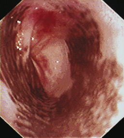
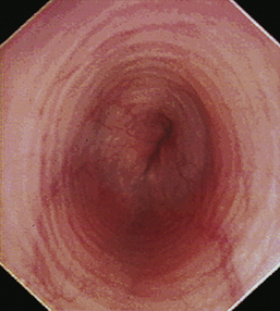
The feline esophagus is composed of striated muscle in the proximal two thirds and smooth muscle in the distal one third, whereas the canine esophagus is composed almost entirely of striated muscle. Longitudinal folds are found throughout the canine esophagus and in the cranial portion of the feline esophagus. In the cat, circumferential mucosal folds in the caudal esophagus form prominent annular ridges that have a herringbone pattern on contrast esophagrams and appear endoscopically as a pattern of circular rings (Figure 3-5). This ringlike appearance is not seen in the dog.


The normal esophageal mucosa in cats and dogs is smooth, glistening, and pale pink or grayish pink in color. It is noticeably less red than the gastric mucosa. Superficial submucosal vessels are normally visible in the feline esophagus (see Figure 3-5) but are not usually seen in the canine esophagus. In heavily pigmented dog breeds such as Chow Chow and Chinese Shar-Pei, the esophagus may contain variably sized confluent gray or black patches of pigmented mucosa (Figures 3-6 and 3-7).


When the GES is first visualized, its configuration should be noted. The lumen of the GES usually forms a slitlike opening that is eccentrically located at the confluence of small radial folds configured in a rosette pattern (Figures 3-8 and 3-9). At the gastroesophageal junction the normal pale pink color of esophageal mucosa changes abruptly to the vivid pink or red color of normal gastric mucosa. In most normal dogs and cats, the GES is closed at the time of endoscopic examination, but this can be affected by anesthetic protocols that decrease the tone of the sphincter and by the degree of air insufflation. In some normal patients the GES can be gaping wide open; however, this is unusual and should raise the suspicion of GES dysfunction, gastroesophageal reflux, or hiatal hernia, especially when the open GES is accompanied by esophagitis and pooling of gastric contents (e.g., food, fluid, or bile) in the esophagus. The GES and cardia should also be examined from the stomach side with the scope in the retroflexed position within the stomach (Figure 3-10); see Chapter 4.


Atlas for Megaesophagus


Megaesophagus is a term that refers to a flaccid dilated esophagus resulting from diffuse hypomotility. Regurgitation associated with impaired esophageal transport of food is the most consistent clinical sign. Weight loss (or poor weight gain) and aspiration pneumonia may be complications. Primary megaesophagus is usually idiopathic and can be either congenital or acquired. Less frequently megaesophagus occurs secondary to other diseases. Congenital idiopathic megaesophagus is characterized by severe dilation of the esophagus and persistent regurgitation of food beginning shortly after weaning. Evidence suggests that the congenital form is inherited in several canine breeds and possibly in Siamese cats. Acquired adult-onset megaesophagus is usually idiopathic, but it also can occur secondary to various underlying neuromuscular diseases that impair esophageal motility, including myasthenia gravis, polymyositis, muscular dystrophies, other polymyopathies, peripheral neuropathies, central nervous system disease, dysautonomia, botulism, tick paralysis, tetanus, anticholinesterase toxicity, lead toxicity, hypoadrenocorticism, and hypothyroidism. Esophageal hypomotility can also occur secondary to esophagitis, hiatal hernia, and obstructing esophageal lesions, such as leiomyomas.


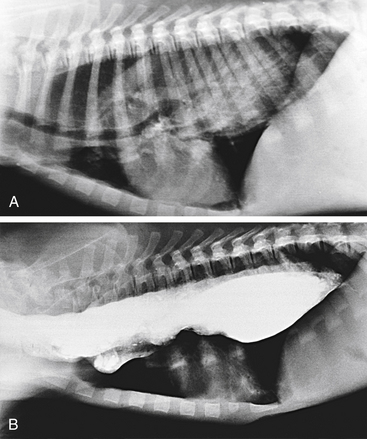
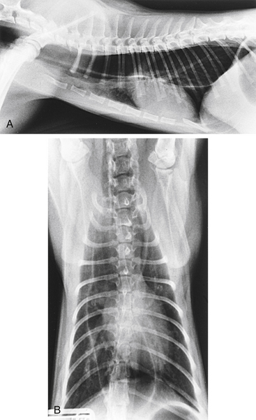
Survey radiography confirms the diagnosis of megaesophagus in most cases and is generally more reliable than endoscopy (Figures 3-11 and 3-12). In normal animals the esophagus is not usually visualized on survey thoracic radiographs, but in megaesophagus the enlarged esophagus distended with air, fluid, or food is readily identified. Disorders of esophageal motility without overt megaesophagus are best characterized by barium swallow videofluoroscopy.


Endoscopic examination is not routinely necessary for confirming megaesophagus and is rarely beneficial for determining the underlying cause; however, esophagoscopy can help rule out obstructive causes of esophageal dilatation (e.g., vascular ring anomalies, strictures, and tumors) and is useful for identifying associated esophagitis that develops in some animals with megaesophagus. Esophagoscopy is not reliable for evaluating motility and the caliber of the esophageal lumen because the normal esophagus becomes flaccid with general anesthesia and dilated after insufflation of air. Furthermore, the esophagus may appear endoscopically normal in animals with mild or focal disturbances of esophageal motility.


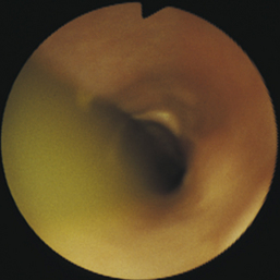
The typical endoscopic appearance of megaesophagus is a markedly dilated, flaccid esophagus extending from the cranial cervical region to the GES, with variable amounts of froth, fluid, and fermenting food residue in the lumen (Figures 3-13 through 3-15). A motility or GES disorder should be suspected when the esophagus contains fluid and ingesta at the start of the examination. Dilation of the entire length of the esophagus distinguishes megaesophagus from segmental dilations or sacculations that develop proximal to luminal obstructions caused by vascular ring anomalies, strictures, tumors, or periesophageal masses. However, diverticulum of the cranial thoracic esophagus is an occasional complication of chronic megaesophagus. The esophageal mucosa in megaesophagus is usually normal in appearance, but secondary esophagitis (mucosal erythema, erosions, and friability) is occasionally observed.


Atlas for Diverticula


Esophageal diverticula are large pouchlike sacculations of the esophageal wall that interfere with the orderly movement of ingesta through the esophagus. Diverticula are relatively uncommon and can be congenital or acquired. Most occur in the cranial mediastinal and epiphrenic regions of the esophagus. Congenital diverticula are caused by abnormalities of embryologic development that result in the herniation of mucosa through a defect in the muscularis. Acquired diverticula can result from external traction and distortion of the esophagus caused by periesophageal inflammatory adhesions (traction diverticula) or most often from increased intraluminal pressure and food impaction (pulsion diverticula) associated with esophageal injury (esophagitis), hypomotility (megaesophagus), or obstruction (e.g., vascular ring anomaly, foreign body, stricture, or tumors). Small diverticula may be of little clinical significance, but larger ones can become impacted with ingesta or hair, causing postprandial distress, retching, odynophagia, and regurgitation. Anorexia, lethargy, and fever may also occur.


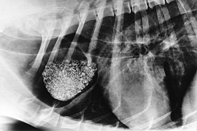
Diverticula can be diagnosed by radiography or endoscopy (Figures 3-16, 3-17, and 3-18). Survey thoracic radiographs show an air-, fluid-, or food-filled mass (pouch) adjacent to the esophagus, and contrast-enhanced radiographs demonstrate filling of the pouch with barium. Esophagoscopy reveals a saclike outpouching from the esophageal lumen, often with erosive esophagitis of the mucosa lining the diverticulum (see Figure 3-17, C). Food, fluid, or hair may have to be removed from the sac before the diverticulum can be adequately visualized (see Figure 3-18). Because of the thin, weakened wall of the diverticular sac, caution must be taken to avoid perforation. Without adequate air distension, the endoscope may enter a blind pouch and be inadvertently forced into the wall. If a diverticulum is small, the only obvious finding may be pooling of fluid. Redundancies in the esophagus that can be mistaken for diverticula are frequently found at the thoracic inlet in clinically normal brachycephalic and Chinese Shar-Pei dogs. Unlike true diverticula, these false diverticula lack associated impaction or esophagitis, and they may decrease or disappear with extension of the neck.


Stay updated, free articles. Join our Telegram channel

Sep 10, 2016 | Posted by in SMALL ANIMAL | Comments Off on Esophagoscopy

Full access? Get Clinical Tree

Get Clinical Tree app for offline access