Laryngoscopy and Tracheobronchoscopy of the Dog and Cat

Chapter 10 Laryngoscopy and Tracheobronchoscopy of the Dog and Cat


Patient Restraint and Positioning


Laryngoscopy can be performed in a matter of minutes. That being said, the patient should nevertheless be examined under a light plane of general anesthesia so that the procedure can be performed in a thorough manner and in a stress-free environment (for the laryngoscopist!). The patient’s plane of anesthesia should be deep enough to allow the oral cavity to be held open without fear that the endoscopist may be bitten, but light enough to retain some degree of normal laryngeal function. An animal that has been properly anesthetized for laryngoscopy still displays a strong gag reflex subsequent to pharyngeal or laryngeal stimulation and will also demonstrate a rapid withdrawal reflex to deep pain. If the patient’s level of anesthesia allows simple gag-free intubation, normal laryngeal motion may be altered because of the anesthesia; this is not ideal, but laryngoscopy should still be performed (see later discussion on the use of doxapram).


Injectable anesthetics are most typically used for laryngoscopy. My preference is propofol used to effect. The dose of propofol needed to generate a proper plane of anesthesia commonly ranges from 1 to 4 mg/kg IV. The drug should be given slowly, over 30 seconds to 2 minutes, to effect. Hypoxemia is absolutely predictable, and every patient should receive supplemental oxygen by mask before and immediately after the procedure. It is also appropriate to intermittently stop laryngoscopy to supplement the patient with oxygen, as needed. Atropine or glycopyrrolate should be used as a premedication to prevent bradyarrhythmias that are sometimes induced by laryngeal manipulation and subsequent vagal stimulation. Topical 1% lidocaine may occasionally be needed to decrease reflex responses (e.g., laryngospasm, swallowing, or gagging) that may be encountered during the procedure. This is much more commonly required in the feline species. Laryngoscopy should be performed with the patient in a sternal recumbent position. A mouth gag should be used to facilitate safe oral examination and to ensure good visualization of the larynx.


Procedure


A thorough laryngoscopic evaluation includes a complete anatomic examination and an assessment of normal laryngeal function and motion. To begin, care should be taken to gently depress the epiglottis from the visual field and gently elevate the soft palate, as needed. Forceful maneuvers can inappropriately fix the laryngeal cartilages in place and artifactually distort otherwise normal laryngeal motion.


Evaluation of Laryngeal Structure


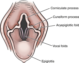
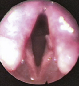
Normal structures that should be evaluated during laryngoscopy include the cricoid, thyroid, and arytenoid cartilages (also the corniculate and cuneiform processes), vestibular folds, vocal folds (cords), laryngeal saccules (lateral ventricles), epiglottis, and aryepiglottic folds (Figure 10-1). Normal mucosa should be pink in color, and superficial vessels may be visible. Secretions are minimal in the area of the normal laryngeal vault. Mucosal hyperemia and edema, excessive secretions, and redundant pharyngeal mucosa are commonly encountered abnormalities. If the lumen of the glottis is smaller than normal, the resulting airflow is turbulent rather than laminar. Turbulent airflow is irritating to mucosa, and this commonly causes at least some of the mucosal erythema and edema that is encountered during laryngoscopy.


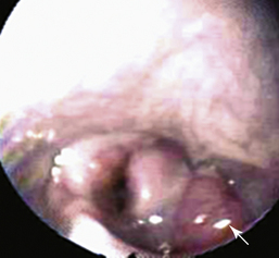
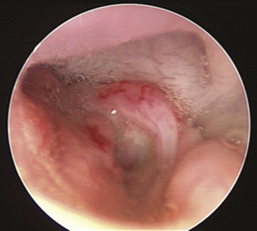
Common structural abnormalities include everted saccules and laryngeal collapse. These abnormalities usually develop as secondary phenomena as a result of more chronic disease and increased negative inspiratory pressures resulting from unilateral or bilateral laryngeal paresis or paralysis. Laryngeal webbing and granuloma formation both typically are secondary to previous surgery or trauma. Both conditions result in stenosis of the glottic lumen, and these fixed lesions often cause severe signs of airway obstruction. Less commonly a pharyngeal or laryngeal rannula, tumor, or foreign body may be found. (Laryngeal abnormalities are depicted in Figures 10-3 through 10-17 in the “Atlas” section). The physical characteristics of secretions or blood should be noted, and the examiner should attempt to determine the anatomic source of the abnormal fluid accumulation. The examiner should recall that secretions (including blood) found in the region of the larynx might have originated in the trachea, lower airways, or lung parenchyma.


Evaluation of Laryngeal Motion


As previously stated, a light plane of anesthesia is most appropriate to properly evaluate normal laryngeal function. However, any level of anesthesia with any routine anesthetic protocol may artifactually distort otherwise normal laryngeal motion. Thus, if laryngeal motion, that is, adduction, abduction, and symmetry of movement, is normal, laryngeal paresis or paralysis may be ruled out. However, if laryngeal motion seems abnormal, doxapram hydrochloride (Dopram-V) 0.5-1.0 mg/kg IV should be administered. Within 30 seconds of administration, this medication increases the rate and depth of respiration, and the increased ventilatory effort persists for a few minutes. Doxapram will not make an abnormal larynx function in a normal manner. Instead, doxapram will override the potentially depressant effects of anesthesia on laryngeal motion. Thus, if the larynx appears abnormal before doxapram and functions normally during doxapram administration, the endoscopist can reasonably assume that laryngeal function is, in fact, normal. Conversely, if laryngeal function remains abnormal after doxapram administration, the endoscopist can reasonably conclude that laryngeal function is abnormal. Side effects of doxapram are minimal and include salivation, muscular tremors (rare), and (uncommonly) vomition.


Laryngeal motion related to vocalization (e.g., whining) or swallowing should not be confused with laryngeal abduction. Normally the size of the glottic lumen is increased on inspiration by an active abduction of the arytenoid cartilages. Complete failure of one or both arytenoid cartilages to abduct (increasing the size of the glottic lumen) is defined as laryngeal paralysis (unilateral or bilateral). Partial failure of movement is referred to as laryngeal paresis. Paradoxical motion (the opposite side is pulled across the midline during inspiration) involving the vocal cord or entire arytenoid cartilage on the affected side is not usually a primary disorder. This is a form of laryngeal collapse and is generally a sequela of chronic laryngeal paresis or paralysis. Laryngeal collapse is a morbid finding because the success of surgery designed to permanently abduct one of the arytenoid cartilages (“lateralization”) is decreased; the “nonlateralized” cartilage and associated vocal fold will tend to move medially, and the glottic opening may not remain in even a semi-open position.


Ancillary Tools and Procedures


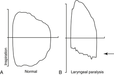
Radiographic evaluation of the larynx does not add significant diagnostic or prognostic information and is not routinely advocated as part of the complete evaluation of laryngeal disorders. A number of investigators have demonstrated that ultrasound of the larynx may be used to diagnose abnormal laryngeal structure or function. However, the practitioner will still need to directly visualize the larynx to make a proper diagnosis. Tidal breathing flow-volume loops (TBFVLs) have been used to document the presence of upper airway obstruction (Figure 10-2). However, an abnormal TBFVL still requires laryngoscopy to confirm the presence and extent of structural and functional laryngeal disorders. A laryngeal electromyogram or biopsy procedure may be used to delineate a specific cause, but these tests are properly performed after the initial laryngoscopic evaluation has been made.


Chest radiographs are usually indicated before laryngoscopy for a few reasons. First, laryngeal disease is commonly a disease of older patients. If surgery to correct abnormalities of the larynx is being considered, radiographic evaluation of the thorax will be valuable in the decision to operate or not. Second, patients with laryngeal disease may be forcefully inspiring against a somewhat closed glottic opening. Interpretation of the lung pattern seen in these patients will be complicated by underinflation and some degree of interstitial bleeding caused by negative inspiratory pressure–induced interstitial capillary rupture. These findings will be valuable in determining the indications for and value and timing of the laryngoscopic procedure.


Complications


A properly performed laryngoscopic examination is a low-risk procedure. However, the clinician should be aware that emergencies might occur during and immediately after laryngoscopy. In a small number of patients, stimulation of the larynx may provoke a very strong vagal response that may result in profound bradyarrhythmias. Administering anticholinergic medication before anesthesia is induced largely prevents this. Laryngeal mucosal hyperemia and edema are commonly encountered in a variety of laryngeal disorders. The manipulation that occurs during laryngoscopy may cause additional mucosal irritation, edema, and, potentially, further narrowing of the airway (a complication that is more common in cats). This is the most important reason why the laryngeal structures should be treated with great respect and should be physically manipulated only as much as is absolutely needed to complete the examination. In addition, some patients with laryngeal disease and upper airway obstruction may have great difficulty maintaining a patent airway during the immediate postanesthetic recovery phase. Patients with upper airway obstruction have adapted a breathing strategy that minimizes discomfort. However, in the immediate postanesthetic recovery phase, a semiaware patient may begin to breathe with increased effort, and this lowers the intraglottic pressures. Lowered intraglottic pressure increases medial movement of the laryngeal cartilages (adduction) and worsens the airway obstruction. A vicious cycle develops in which increased airway obstruction causes panic, increased effort to breathe, and further worsening of airway obstruction. Therefore endotracheal and tracheostomy tubes and instruments should be available (in suspected cases). If a surgically correctable disorder of the larynx is suspected (e.g., laryngeal paralysis), the laryngoscopic examination should be scheduled so that surgical correction can immediately follow the examination, thereby avoiding a second anesthetic procedure.


Differences Between Feline and Canine Laryngoscopy


The laryngeal vault of dogs and cats are anatomically, grossly different. Specifically, the corniculate and cuneiform processes of the arytenoid cartilages are well developed and are much more prominent in dogs than in cats. Furthermore, mucosa lining the feline laryngeal vault becomes edematous after manipulations that cause no edema in canine patients. This edematous response may be dramatic and can result in serious airway obstruction (see Figure 10-5 in the “Atlas” section). The feline larynx may respond to light touch with spasm, further occluding the upper airway. For these reasons it is critical to place a few drops of 1% lidocaine on the surface of the feline laryngeal structures before laryngoscopy is performed. It is also important for the operator to be very gentle when manipulating this area.


Atlas for Laryngoscopy Pages 334-338
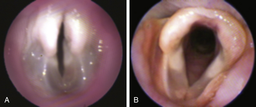
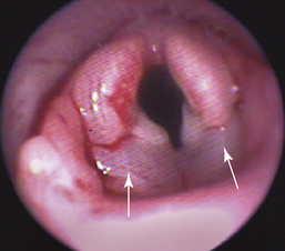
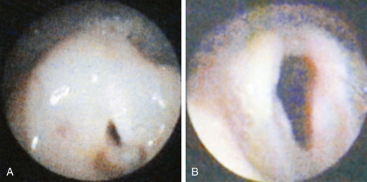
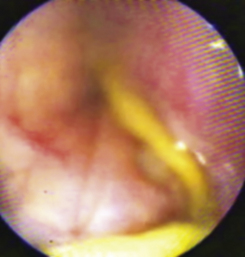
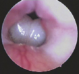
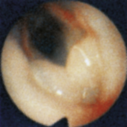
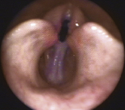
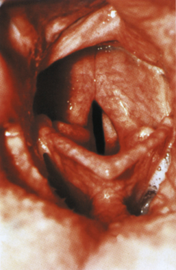
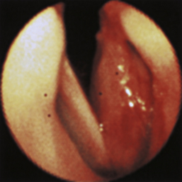
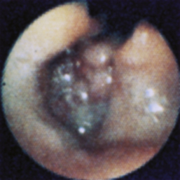
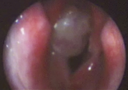


Laryngeal Edema


Figure 10-3, p. 334, Laryngeal edema in a cat

Figure 10-4, p. 334, Laryngeal edema in a cat with smoke inhalation injury

Figure 10-5, p. 335, Severe laryngeal edema in a cat due to traumatic intubation

Laryngeal Foreign Bodies


Figure 10-6, p. 335, String foreign body in a dog with acute gagging

Laryngeal Sacculitis


Figure 10-7, p. 335, Everted laryngeal saccules in a dog with noisy breathing

Figure 10-8, p. 335, Saccular edema and eversion in a cat

Laryngeal Collapse


Figure 10-9, p. 336, Laryngeal collapse in a dog with exercise intolerance

Figure 10-10, p. 336, Partial laryngeal collapse in a dog with noisy breathing

Figure 10-11, p. 336, Laryngeal collapse in a dog with exercise intolerance and loss of bark

Nonneoplastic Growths


Figure 10-12, p. 337, Vocal fold granuloma in a dog

Figure 10-13, p. 337, Laryngeal granuloma in a dog

Figure 10-14, p. 337, Vocal fold tear in a dog

Laryngeal Neoplasia


Figure 10-15, p. 338, Undifferentiated carcinoma in a cat

Figure 10-16, p. 338, Lymphoma in a cat

Figure 10-17, p. 338, Squamous cell carcinoma in a dog

Figure 10-18, p. 338, Thyroid carcinoma causing significant airway obstruction in a dog


Tracheobronchoscopy of the Dog and Cat


In 1904 Dr. Chevalier Jackson developed the first rigid bronchoscope; however, it took 64 more years (1968) before fiberoptic bronchoscopy became part of clinical pulmonary medical practice in human medicine. Over the past 20 years, respiratory endoscopy has also become an important diagnostic technique in the practice of high-quality veterinary medicine. Before the development of the bronchoscope, veterinary patients with clinical signs referable to the respiratory system were evaluated with the techniques of chest auscultation, thoracic radiography, and transtracheal washing. Bronchoscopy advanced our ability to examine these patients; with a bronchoscope we can better understand the nature, vascularity, extent, and distribution of many pathologic changes in airway wall structure and function. A microbiology brush can be easily passed into areas of focal infection without contamination of “healthy” sites, which avoids a common complication that can occur during unguided transtracheal or bronchopulmonary washing through an endotracheal tube. Exfoliated cells from primary or metastatic neoplastic lesions can be identified with the technique of bronchoalveolar lavage, which has made it possible to stage and treat cancer more effectively. Left atrial compression of a mainstem bronchus can be identified, which can help confirm the presence of cardiomegaly as a cause of coughing and can help distinguish cardiac- and respiratory-related disorders. The general experience of most clinicians who use bronchoscopy in their practices of veterinary medicine and surgery can be summarized in one line: “How did we ever get along without it?”


Indications


The two most frequent indications for performing bronchoscopy in veterinary patients are (1) acute or chronic cough that is unanticipated or unresponsive to standard medical therapy and (2) unexplained radiographic infiltrates (Box 10-1). Bronchoscopy can also be invaluable in distinguishing cardiac from respiratory causes of a cough and for staging metastatic lung cancer. Tracheobronchoscopy should also be considered in veterinary patients with noisy breathing or stridor for which laryngoscopy fails to confirm the cause.


Bronchoscopy is particularly valuable in collecting samples for microbiological, cytologic, and histologic analysis. Tracheobronchial biopsy is primarily performed to obtain a histologic diagnosis for abnormal endobronchial growths or parenchymal infiltrates. Microbiological sampling of infected areas within the respiratory tree is greatly assisted by bronchoscopic guidance because the sampling brush can be directed into specific areas of interest. This technique offers the best chance of retrieving diagnostic samples and minimizes the risk of cross-contamination of less involved sites, as can occur when nonspecific washing techniques are used. Bronchoalveolar lavage is a technique that can be used to assess the morphology and distribution of cells that line individual lung segments, and the retrieved samples can be processed to include total and differential counts of the cells obtained.


Bronchoscopy itself has no specific contraindications. However, the procedure should not be performed in patients that are not candidates for general anesthesia. Importantly, all standard bronchoscopes are larger in diameter than standard endotracheal tubes used for cats; therefore bronchoscopy in the feline patient must be done with great speed and skill to avoid predictable hypoxemia and respiratory compromise.

Stay updated, free articles. Join our Telegram channel

Sep 10, 2016 | Posted by in SMALL ANIMAL | Comments Off on Laryngoscopy and Tracheobronchoscopy of the Dog and Cat

Full access? Get Clinical Tree

Get Clinical Tree app for offline access