57 Episcleritis INITIAL PRESENTATION Abnormal appearance – red PRESENTING SIGNS Episcleritis is seen in dogs rather than in cats and the animal will be presented because of a red eye – usually affecting only one eye, but sometimes both. Often the owner thinks that the dog has conjunctivitis. The condition is not usually painful and the owner reports no apparent visual disturbance. CASE HISTORY In most cases there is no previous history of ocular disease or relevant systemic disease. The condition is usually insidious in onset, such that there has been some intermittent ocular redness for days to weeks before the animal is brought to the surgery – the owner may notice that this is worsening. Usually there is minimal discomfort – perhaps mild blepharospasm only – and the owner will not be worried by this. CLINICAL EXAMINATION 
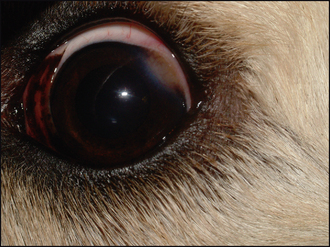
General clinical examination is unremarkable. On ophthalmic examination the most obvious abnormality is the red eye. Marked episcleral congestion will be present, together with some overlying conjunctival hyperaemia. Thus the superficial conjunctival vessels – the thin bright red branching ones – will be engorged, while the deeper, darker, straighter episcleral vessels will be thickened and more tortuous than usual. There will be some diffuse swelling in the area but this will not be excessive (not true chemosis) (Figure 57.1). Figure 57.1 Mild episcleritis in a golden retriever. Note the slight episcleral swelling and hyperaemia with corneal vascularization and corneal oedema. The episcleral congestion can be diffuse, affecting large quadrants of the globe, or can be a discrete nodular swelling. With the latter, the lesion is not usually firmly attached to the underlying sclera and can be moved slightly with a moistened cotton bud. However, if the nodule extends into the cornea it will be less mobile. Some peripheral corneal oedema will be present, giving a blue tinge to the cornea adjacent to the limbus. This will be denser and affect slightly more of the cornea where the episcleral involvement is more severe. Fluorescein testing is negative. The eye will be visual (unless very advanced disease is present involving the posterior segment but this is unusual) with a normal menace response, pupillary light reflexes and no evidence of intraocular disease. Intraocular pressures will be normal. In most cases fundus examination is normal, but in very severe cases there might be evidence of active chorioretinitis (with fuzzy, grey retinal elevations and perivascular cuffing as a direct extension from the outer episcleral inflammation) and even optic neuritis in a few cases. DIFFERENTIAL DIAGNOSES • Conjunctivitis • Chronic superficial keratitis • Uveitis • Panophthalmitis • Healing peripheral corneal ulcer • Glaucoma • Limbal neoplasia (for nodular form) • Retrobulbar cellulitis CASE WORK-UP The condition can usually be diagnosed on clinical examination alone. However, in some patients, particularly if suffering from the nodular form of the disease, biopsy is recommended to differentiate from neoplastic disease. This requires heavy sedation or general anaesthesia, and the biopsy should be deep enough to include the episcleral tissue, not just the conjunctiva (it is for this reason that general anaesthesia is advised). NURSING ASPECTS Apart from treating with topical and sometimes systemic medication, there is no specific nursing required with cases of episcleritis. Only gold members can continue reading. Log In or Register to continue You may also needBlepharitisDeep corneal ulcerRabbit uveitisFeline glaucomaCanine intraocular neoplasiaAberrant conjunctival overgrowth in rabbitsSecondary canine glaucomaCanine lens luxation Share this:Click to share on Twitter (Opens in new window)Click to share on Facebook (Opens in new window)Click to share on Google+ (Opens in new window) Related Tags: Saunders Solutions in Veterinary Practice Small Animal Ophthalmo Sep 10, 2016 | Posted by admin in SMALL ANIMAL | Comments Off on Episcleritis