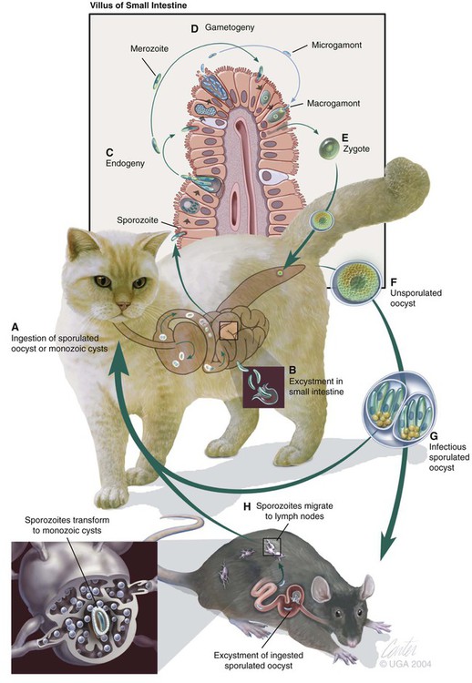
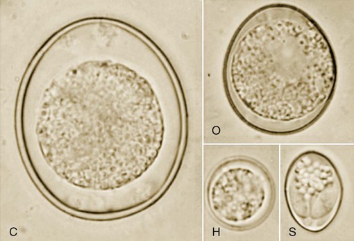
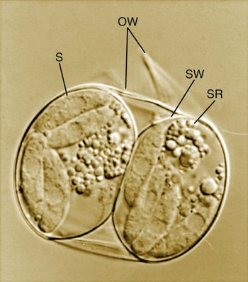
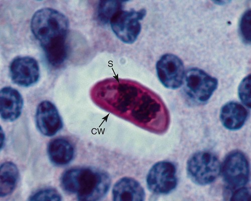
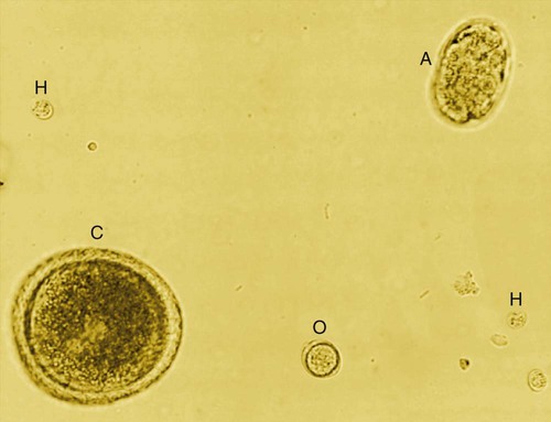
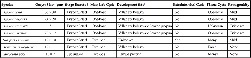
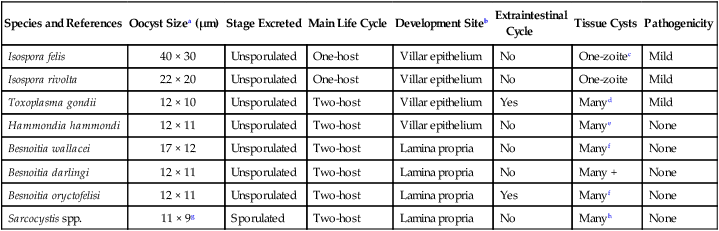
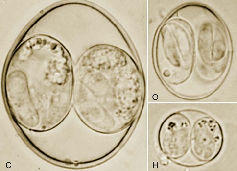
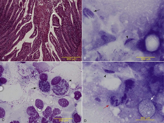
Coccidia are obligate intracellular parasites normally found in the intestinal tract. They belong to the phylum Apicomplexa, class Sporozoasida, order Eucoccidiorida, and, depending on the species, family Eimeriidae, Cryptosporidiidae, or Sarcocystidae. Coccidian genera that infect cats and dogs are Isospora (also called Cystoisospora), Hammondia, Besnoitia, Sarcocystis, Caryospora, Toxoplasma, Neospora, Cryptosporidium, and Cyclospora species.13 Most of the genera are covered in this chapter; however, Toxoplasma and Neospora are in Chapter 79 and Cryptosporidium and Cyclospora are in Chapter 81. Another coccidian genus, Eimeria, found commonly in herbivores, birds, lagomorphs, and rodents, is found only in feces of dogs and cats after they ingest intestinal contents or feces from these animals. The oocysts pass unchanged through the feline or canine intestine. Some coccidians of dogs remain unclassified. Intestinal coccidia discussed in this chapter are host specific (Tables 80-1 and 80-2). Infections of definitive or intermediate hosts generally only occur in cycles established by evolution. Some aberrant cycles exist, such as with Sarcocystis neurona infections in horses. Human health risks from these parasites are considered minimal to nonexistent, even in immunosuppressed humans. TABLE 80-1 Summary of Biology of Coccidia of Dogs ?, Oocysts are considered to be the same size as I. ohioensis but were not described. aAverage size of unsporulated oocyst. bSchizonts in the small intestine of the dog. cThese cysts contain 1 sporozoite and have been found only in experimentally-infected animals fed oocysts. dTissue cysts are microscopic, contain many bradyzoites and are found in the central nervous system and muscles. eTissue cysts are not confirmed. gSarcocystis cysts are often macroscopic and occur only in the intermediate hosts. From Ref. 28. TABLE 80-2 Summary of Biology of Coccidia of Cats aAverage size of unsporulated oocyst. bSchizonts in the small intestine of the cat. cThese cysts contain one sporozoite and have been found only in experimentally infected animals fed oocysts. dTissue cysts are microscopic and contain many bradyzoites in almost all tissues of the cat. eTissue cysts are not found in the cat. They are found mainly in muscles of rodents fed H. hammondi oocysts. fBesnoitia cysts are found only in the intermediate hosts and can be macroscopic. hSarcocystis cysts (sarcocysts)are often macroscopic and occur only in the intermediate host. From Ref. 28. All coccidians have an asexual and a sexual cycle. In some genera, such as Sarcocystis, the asexual and sexual cycles occur in different hosts, whereas in Isospora both cycles may occur in the same host (see Tables 80-1 and 80-2 and Fig. 80-1). The oocyst is the environmentally resistant stage in the life cycle of all coccidia and is excreted in feces of the definitive host. A representative coccidian life cycle is best described as follows. Oocysts are passed unsporulated in feces and contain a single nucleated mass called a sporont, which almost fills the oocyst (Fig. 80-2). After exposure to warm (20° C to 37° C [68° F to 98.6° F]) environmental temperatures and moisture, oocysts sporulate, forming two sporocysts. Within each sporocyst are four sporozoites (Fig. 80-3). The sporozoites have a banana shape and are the infective stage (Fig. 80-4). They can survive environmental exposure, protected inside the oocysts for many months. After ingestion of sporulated oocysts by cats or dogs, sporozoites excyst in the intestinal lumen, and the sporozoites initiate the formation of schizonts or meronts (Fig. 80-5). During schizogony or merogony, the sporozoite nucleus divides into two, three, or more nuclei, depending on the parasite and the stage of the cycle. After nuclear division, each nucleus is surrounded by cytoplasm, forming a merozoite. The number of merozoites within a schizont varies from two to several hundred, depending on the stage of the cycle and the species of coccidia. Merozoites are released from the schizont when the host cell ruptures. The number of schizogonic cycles varies with the parasitic species. First-generation merozoites repeat the asexual cycle and form second-generation schizonts or transform into microgamonts (males) and macrogamonts (females). The microgamont divides into many tiny microgametes. A microgamete fertilizes a macrogamete, and an oocyst wall is formed around the zygote. The life cycle is completed when unsporulated oocysts are excreted in feces (see Fig. 80-1). The prevalence rate of coccidial infections in dogs and cats varies with factors such as climate, age, and housing of animals. Geographic prevalence studies have been done in many countries under a variety of housing conditions for dogs or cats.* In one study in the United States, involving more than 600,000 client-owned cats evaluated at veterinary practices, those at increased risk were under 4 years of age, purebred, and living in the East, South, Central, and Mountain regions.12 Members of the genus Isospora, the most commonly recognized coccidians infecting dogs or cats, are species specific for the definitive host. At least four species—Isospora canis, Isospora ohioensis, Isospora burrowsi, and Isospora neorivolta—infect dogs (see Table 80-1), and two species—Isospora felis and Isospora rivolta—infect cats (see Table 80-2). The life cycles of Isospora infecting dogs and cats is similar to the basic coccidian intestinal cycle, except an asexual cycle can also occur in the definitive or intermediate host. On ingestion by definitive or suitable paratenic (intermediate) hosts, oocysts excyst in the presence of bile, and free sporozoites invade the intestine. Some sporozoites penetrate the intestinal wall and enter mesenteric lymph nodes or other extraintestinal tissues (e.g., spleen and liver), where they form enlarging unicellular cysts (Fig. 80-6). If no replication occurs, the host is called a paratenic host rather than an intermediate host. Monozoic cysts of Isospora may remain in extraintestinal tissues of definitive and paratenic hosts for the life of the host. In cell culture, these monozoic cysts have been shown to contain a single sporozoite.59 In dogs and cats, these cysts may serve as a source of intestinal reinfection and relapse of enteric coccidiosis. Ingestion of monozoic cysts in paratenic hosts leads to intestinal infection in the definitive dog and cat host. The life cycle after the ingestion of paratenic host is the same as after the ingestion of sporulated oocysts from feces. Diarrhea with coccidiosis in immunocompetent animals probably represents incidental or concurrent infections with coccidia and other infectious agents because coccidial infection can be present in the absence of clinical illness. Enzootic infections are frequently found in catteries or kennels where animals congregate. Clinical signs are most apparent in neonates. Experimental studies have shown that clinical signs of intestinal disease are uncommon unless large numbers of oocysts are fed to very young (younger than 1 month) or immunosuppressed animals.4 Infections with I. canis produce longer prepatent periods, longer excretion times, and higher concentrations of oocysts as compared to infections with I. ohioensis.4 However, diarrhea did develop in 8-week-old pups experimentally infected with I. canis, even in the absence of immunosuppression or concurrent infections.59 Clinically, severe diarrhea has been associated with naturally occurring coccidiosis in immunosuppressed dogs and cats or in those with other co-infections or co-infestations.45,50 In some cases, the enteric coccidial infections are incidental to other infecting organisms or causes of diarrhea.67 German shepherd dogs may have an increased susceptibility to clinical infection.40,66 Diarrhea with weight loss and dehydration and, although rare, hemorrhage is the primary sign attributed to coccidiosis in dogs and cats. Anorexia, vomiting, mental depression, and ultimately death may be seen in severely affected animals. Severely immunosuppressed dogs and cats may have extraintestinal stages in macrophages of the lymphocyte-depleted mesenteric lymph nodes or extraintestinal tissues. Intestinal coccidial infection in dogs and cats is diagnosed by identification of the oocysts with any of the fecal flotation methods commonly used to diagnose parasitic infections (see Examination of Feces, Chapter 70). Shedding of oocysts by some animals may be erratic; therefore, repeated examinations are recommended. In dogs, only I. canis can be identified with certainty by oocyst size and shape (see Fig. 80-2). The two species of Isospora found in cats can be readily distinguished by oocyst size (see Fig. 79-14). Oocysts of I. felis in cats and I. canis in dogs are large and easily distinguished from small oocysts, whereas it is almost impossible to distinguish I. rivolta, I. burrowsi, and I. ohioensis morphologically (Fig. 80-7; see also Figs. 80-2 and 80-3). Oocysts of Hammondia heydorni and Neospora caninum are very difficult to distinguish from each other (see Chapter 79).24,76 Although I. felis-, I. rivolta-, I. canis-, and I. ohioensis-like oocysts are passed unsporulated in freshly excreted feces, they sporulate partially by the time a fecal examination is made. Partially sporulated oocysts contain two sporocysts without sporozoites. Isospora spp. may sporulate within 8 hours of excretion, and these Isospora are highly infectious. In severe cases, merozoites and schizonts from the small intestine maybe found in smears of diarrheic feces (see Fig. 80-5). Specific therapy involves the use of drugs that are coccidiostatic rather than curative (Table 80-3). However, as with many protozoal diseases, the presence of low-level infection may lead to premunition. This is a state of chronic low-level persistent infection, which leads to resistance to further infection or reinfection, thereby preventing clinical illness. The drugs shorten the prepatent period and may shorten the course of the disease. TABLE 80-3 Anticoccidial Drugs for Dogs and Cats
Enteric Coccidiosis
Species
Oocyst Sizea (µm)
Stage Excreted
Main Life Cycle
Development Siteb
Extraintestinal Cycle
Tissue Cysts
Pathogenicity
Isospora canis
38 × 30
Unsporulated
One-host
Villar epithelium
No
One-zoitec
Mild
Isospora ohioensis
24 × 20
Unsporulated
One-host
Villar epithelium
No
One-zoite
Mild
Isospora neorivolta
?
Unsporulated
One-host
Villar epithelium and lamina propria
No
Unknown
Unknown
Isospora burrowsi
20 × 17
Unsporulated
One-host
Villar epithelium and lamina propria
No
One-zoite
Unknown
Neospora caninum
12 × 10
Unsporulated
Two-host
Unknown
Yes
Manyd
Mild
Hammondia heydorni
12 × 11
Unsporulated
Two-host
Villar epithelium
No
Raree
None
Sarcocystis spp.
11 × 9f
Sporulated
Two-host
Lamina propria
No
Manyg
None
Species and References
Oocyst Sizea (µm)
Stage Excreted
Main Life Cycle
Development Siteb
Extraintestinal
Cycle
Tissue Cysts
Pathogenicity
Isospora felis
40 × 30
Unsporulated
One-host
Villar epithelium
No
One-zoitec
Mild
Isospora rivolta
22 × 20
Unsporulated
One-host
Villar epithelium
No
One-zoite
Mild
Toxoplasma gondii
12 × 10
Unsporulated
Two-host
Villar epithelium
Yes
Manyd
Mild
Hammondia hammondi
12 × 11
Unsporulated
Two-host
Villar epithelium
No
Manye
None
Besnoitia wallacei
17 × 12
Unsporulated
Two-host
Lamina propria
No
Manyf
None
Besnoitia darlingi
12 × 11
Unsporulated
Two-host
Lamina propria
No
Many +
None
Besnoitia oryctofelisi
12 × 11
Unsporulated
Two-host
Lamina propria
Yes
Manyf
None
Sarcocystis spp.
11 × 9g
Sporulated
Two-host
Lamina propria
No
Manyh
None
Intestinal Coccidiosis
Isospora Species
Epidemiology
Clinical Findings
Diagnosis
Therapy
Druga
Species
Dose (mg/kg)b
Route
Interval (hours)
Duration (days)
Sulfadimethoxinec
B
50–60
PO
24
5–20
Sulfaguanidine
B
100–200
PO
8
5
Trimethoprim-sulfonamide
D
30–60d
PO, SC
24
5
B
15–30e
PO, SC
12–24
5
Ormetoprim-sulfadimethoxine
D
66f
PO
24
7–23
Furazolidoneg
B
8–20
PO
12–24
5
Amprolium
D
300–400 (total)h
PO
24
5
D
110–200 (total)i
PO
24
7–12
C
60–100 (total)
PO
24
7
Quinacrine
B
10
PO
24
5
Spiramycin
H
50–100 (total)j
PO
24
5
Clindamycin
C
10
PO, SC, IM
12
7-28
Toltrazuril
D
15–30
PO
24
1–6k
Diclazuril
C
25
PO
24
1
Ponazuril
D
30–50l
PO
24
1–7
C
15m
PO
24
7
Roxithromycin
H
2.5
PO
12
15 ![]()
Stay updated, free articles. Join our Telegram channel

Full access? Get Clinical Tree