54 Enophthalmos
CLINICAL EXAMINATION
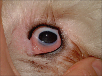
On ophthalmic examination the first thing to determine is whether the eye is truly enophthalmic, i.e. sunken back into the socket, or whether it is just smaller than it should be. Comparison of the two eyes will assist, as will viewing the animal from above as well as from directly in front. A small eye can be present in young animals; sometimes it will be normally developed and visual, but just smaller than expected, but other ocular abnormalities, such as cataract, nystagmus and persistent pupillary membrane, might be present (Figure 54.1).
An acquired small globe develops following severe intraocular damage and is termed phthisis bulbi. It can follow severe trauma, uveitis or chronic glaucoma (Figure 54.2). A severely phthitic eye will usually be blind and grossly abnormal, with conjunctival hyperaemia, episcleral congestion and frequently severe keratitis such that detailed intraocular examination is not possible. Attempts at measuring intraocular pressure in such patients will be difficult since the globe becomes very soft such that obtaining accurate readings is problematical.
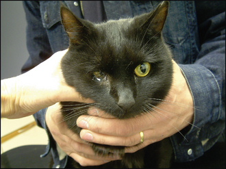
The patient should be examined for evidence of ocular pain which can cause retraction of the globe. Thus a corneal lesion such as an ulcer or laceration, and severe uveitis or a lens luxation can all cause sufficient ocular pain to contract the retractor oculi muscle such that the globe sinks and the nictitans membrane becomes elevated. An ocular discharge is likely to be present in painful eyes – varying from serous epiphora to overt purulent material. This should be differentiated from the accumulation of mucus seen at the medial canthus in patients with deep set eyes (see Case example 12.1 on medial canthal pocket syndrome). Pupil size should be checked – a miotic pupil in an enophthalmic eye should suggest Horner’s syndrome, especially if the eye is not inflamed with no evidence of uveitis. The patient should be examined in bright and dim light, and the degree of anisocoria assessed. It will be more noticeable in the dark, as the affected pupil fails to dilate. The other signs of Horner’s syndrome, namely ptosis and elevation of the nictitans membrane, will usually be present, together with the enophthalmos and miosis, and all result from damage to the sympathetic supply to the eye.


