CHAPTER 8 Dry-Mount Fecal Cytology
There are several diagnostic tests of feces that may be necessary during complete evaluation of patients with clinical signs referable to the gastrointestinal tract. Optimal fecal assessment, including potential tests (e.g., wet-mount fecal cytology, dry-mount fecal cytology, bacterial culture, fecal antigen detection methods, fecal flotation, fecal sedimentation, Baermann technique) and their required sample handling, diagnostic indications, and interpretations, have been compiled elsewhere (Broussard, 2003). Dry-mount fecal cytology (air-dried smear) is one component of a thorough diagnostic evaluation of patients with gastrointestinal signs. Since some fecal pathogens can be morphologically indistinguishable from incidental nonpathogens, results of dry-mount fecal cytology should be interpreted in the context of the patient’s clinical presentation and results of other diagnostic tests, such as wet-mount fecal cytology, bacterial culture, and fecal antigen detection methods.
When evaluating dry-mount fecal cytology, it may be useful to apply a systematic approach aimed at evaluation of attendant background cells and detection of abnormal eucaryotic cells and pathogenic microorganisms (Box 8-1).
BOX 8-1 Guidelines for Systematic Method for Dry-Mount Fecal Cytology Evaluation
SAMPLE COLLECTION AND PROCESSING
The method of fecal sample collection can affect which portion of the rectum is represented (luminal vs. mucosal) and the cellular (eucaryotic and procaryotic) content of the sample. The ideal sampling method, desired amount to collect, and subsequent sample handling depends upon the intended fecal testing (Broussard, 2003). Important goals of sampling for fecal cytology include examination of a fresh sample (less than 5 minutes old) that is representative of the mucosal surface and preparation of a fecal thin film preparation that is not excessively dense. Excessively dense areas of fecal thin film preparations are prone to detach during staining or, if they do not fall off during staining, obscure cytologic details. Because the cellular content of fecal material is dynamic and will likely continue to change after sample collection, immediate examination following collection is recommended.
For fecal cytology, there are a few possible sampling methods, including collection of unadulterated fecal material, rectal saline lavage, and rectal scraping. A small amount of fresh fecal material may be obtained during digital rectal examination or using a moistened, cotton-tipped applicator or fecal loop if digital rectal exam is not possible. Immediately collected, voided feces may also be used; however, voided samples may be more representative of the luminal portion of the rectum, which is not ideal for fecal cytology. Voided fecal material is a preferred sample for other diagnostic tests, such as fecal flotation, fecal sedimentation, or the Baermann technique, since it is more copious and may be rich in parasitic eggs and cysts (Broussard, 2003). Usually, if a small amount of fecal material is obtained using a moistened, cotton-tipped applicator, the cotton tip of the applicator can be gently rolled on the slide to prepare a direct thin film. Otherwise, slight dilution of the feces may be used to prepare a thin film preparation by placing a drop of sterile, normal saline on a clean, glass microscope slide, adding a very small amount of fecal material (no larger than a match-head), mixing with a sterile, wooden applicator, and spreading as for other fluid thin film preparations. Prior to and after spreading, newsprint should be legible though the preparation.
Rectal saline lavage enriches for mucosal material, including mucus, motile protozoa, and bacteria. Lavage fluid contains relatively less luminal fecal material and is ideal for fecal cytology (Broussard, 2003). Properly collected lavage fluid should have a mudlike consistency, and a single drop can be used to prepare a thin film preparation.
NORMAL OR INCIDENTAL MICROSCOPIC FINDINGS
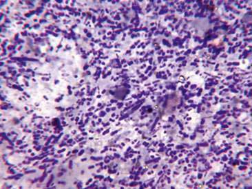
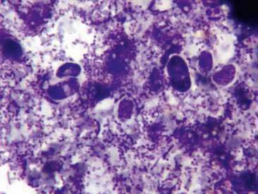
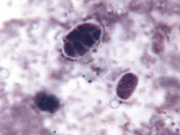
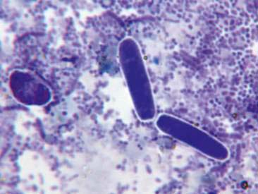
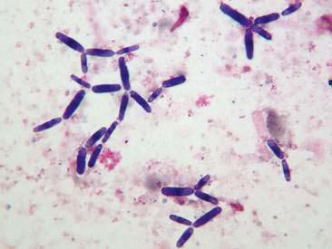
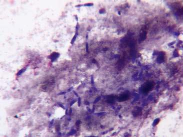
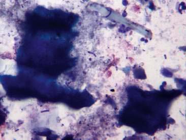
The background microbial flora should be predominantly comprised of an extremely polymorphic population of several different bacterial bacilli (Fig. 8-1). There should be fewer than 5 spore-forming bacilli per 100× objective field (Broussard, 2003). Cocci should be absent or only rarely observed. A low number of extracellular, circular or ovoid, 5 to 10 μm diameter, stippled, variably basophilic yeast with a thin, colorless capsule may occasionally be seen (Figs. 8-2, 8-3, 8-4). While these yeast structures are frequently found in diarrheal stools, it is not certain whether there is a direct causal relationship.
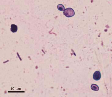
Cyniclomyces guttulatus (formerly Saccharomycopsis guttulata) is part of the normal flora of some rodents and lagomorphs (rabbits, guinea pigs, and chinchillas) (Zierdt, 1988). Occasionally, a low number of individual or doublet Cyniclomyces may be observed as an incidental finding in canine feces and are thought to represent a nonpathogenic result of coprophagia (Fig. 8-5). However, there is uncertainty about whether this yeast is entirely nonpathogenic in dogs. A few preliminary research or anecdotal reports have associated this yeast with clinical cases of chronic diarrhea (Houwers and Blankenstein, 2001; Mandigers, 2007; Saito et al., 2000). During evaluation of some patients with chronic diarrhea, the only abnormality detected in dry-mount fecal cytology may be the presence of an extremely large number of Cyniclomyces both individually and in large mats of numerous budding organisms (Figs. 8-6, 8-7). The observation of many apparently replicating organisms in a fresh fecal sample may represent a factor contributing to the ongoing diarrhea or may simply represent abnormal flora due to underlying disease, physiologic processes, or prior antimicrobial treatments. At this time, the pathogenicity of this yeast is unproven, though it is occasionally observed in large number in the feces of dogs with chronic diarrhea; in such cases, this observation should be reported as an abnormal finding.
It is common to see a variable amount of irregularly shaped, colorless or amber material and green-blue material with parallel cell walls, representing digesta and ingested plant matter, respectively (Fig. 8-8). The background also commonly contains a variable amount of amorphous basophilic material consistent with mucus. The amount of mucus observed depends on whether the underlying disease is associated with rectal oversecretion of mucus and whether the sampling method is more representative of the rectal mucosa or the rectal lumen. Being more representative of the rectal mucosa, samples obtained by rectal saline lavage or rectal scraping may contain more mucus than samples collected by other means.
< div class='tao-gold-member'>
Stay updated, free articles. Join our Telegram channel

Full access? Get Clinical Tree


