Drugs Used in Ophthalmic and Otic Disorders
Learning Objectives
After studying this chapter, you should be able to
1. Describe the clinical indications for common ophthalmic and otic agents.
2. Describe the products used to diagnose ocular problems.
3. Identify the different classes of ophthalmic and otic agents.
4. Identify the possible adverse reactions and contraindications of many commonly used agents.
Key Terms
Blepharospasm
Cerumen
Conjunctivitis
Cycloplegia
Distichia (distichiasis)
Ectropion
Entropion
Glaucoma
Horner’s syndrome
Hyphema
Intracameral injection
Keratitis
MRSA
Mydriasis
Open-angle glaucoma
Otoacariasis
Uvea
Uveitis
Introduction
The sense of smell is highly developed in animals, but the sense of sight also plays an important role in an animal’s health and well-being. Cats rely on excellent eyesight because they are animals of prey. This prey trait can provide cat owners with much laughter as a string or a feather toy is pulled around the house. Horses rely on good eyesight to perform their best in equestrian events. Police dogs, hunting dogs, seeing-eye dogs, and herd dogs rely on their eyesight to interpret hand signals when working in the field.
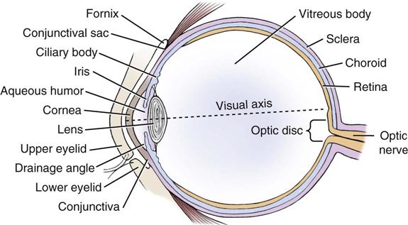
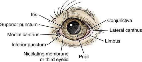
An ophthalmic examination includes the use of diagnostic agents to determine any ocular problems that may be present in the patient. Additionally, an examination of the ocular features (eyelids, eyelashes, sclera, cornea, third eyelid [i.e., nictitating membrane]), pupil, anterior chamber, iris, and lens), all of which can be seen without highly specialized equipment (McCurnin, 2006), are also important to evaluate. Some dog breeds (shar-pei, cocker spaniels, English bulldogs, and others) are genetically predisposed to conditions that may require cosmetic surgery to correct faults. Three of these conditions are known as entropion, ectropion, and distichiasis.
Topical administration of eye drops or ointment is the most common method of treatment involving disorders of the eye. It is the veterinary technician’s duty to educate the client by demonstrating the proper way to administer eye medication. Products for ocular treatment are usually available as solutions or ointments. Drug penetration is one factor that veterinarians must consider when choosing a topical ophthalmic agent. Topical agents are more readily absorbed into the anterior chamber than the posterior chamber. For this reason, these agents have limited use in posterior eye disorders. Systemic agents may be more effective. Lipid-soluble agents readily penetrate the corneal epithelium and endothelium layers. Water-soluble agents readily penetrate the corneal stroma layer (Figures 10-1 and 10-2). Most topical ophthalmic medications require several applications per day because the eye continuously secretes tears that wash away the medication. Ointments tend to necessitate less frequent applications than drops. However, ointments may blur an animal’s vision for a short period after application. Recently, some clinicians believe that ophthalmic drops may be more effective than ointments.
Client education is invaluable for proper treatment of an eye disorder. It is important that clients understand that the applicator tip of the drug’s container should not touch the eye’s surface or the conjunctiva because bacteria from the patient’s eye may contaminate the remaining drug contents inside the container. This is also important to remember when veterinary personnel are treating several patients in a veterinary hospital with the same drug container.
Clients placing telephone calls to the veterinary hospital to discuss a potential eye problem in a companion animal should be made to realize that these situations may be considered an emergency. Unfortunately, some clients tend to let an ocular problem progress to severe stages before treatment is sought. Veterinary technicians should remind clients that animals have only two eyes and the importance of vision should not be minimized.
Diagnostic Agents
The use of diagnostic agents to determine what problems may exist with the eyes is an important part of the ophthalmic examination. Some diagnostic agents should be used before others so that results are not misinterpreted.
 Technician Notes
The Schirmer tear test is one of the first diagnostic procedures to be employed so that tear evaluation is calculated correctly.
 Technician Notes
If a bacterial culture is indicated, the culture should be obtained before cleaning the eye or administering other ophthalmic drugs. Therefore, an eye examination should be carried out in a methodical manner in order for a proper diagnosis to be made.
Fluorescein Sodium
Clinical Uses
Fluorescein stain is commonly used to stain the patient’s cornea in cases in which a corneal ulcer is suspected. The stain fluoresces, and if a corneal ulcer is present it can be observed with the use of a cobalt blue filter and a light source. The corneal epithelium will not stain because the corneal stroma, which is a lipid membrane, repels the stain. Fluorescein stain is also used to diagnose patency of the nasolacrimal outflow duct.
Dosage Forms
Fluorescein stain is manufactured on paper strips.
 Technician Notes
Lissamine Green
Clinical Uses
Lissamine green is used to diagnose corneal damage and to quantify tear production. This product does not sting like Rose Bengal, but interpretation requires more experience. Fluorescein stain is a more reliable indicator of corneal damage.
Dosage Form
Adverse Side Effects
Side effects are uncommon.
Phenol Red Thread
Clinical Uses
This product is used to evaluate tear production. Phenol red thread is a new accurate way to measure tear production as compared with the Schirmer tear test.
Dosage Form
Adverse Side Effects
Side effects are uncommon.
Rose Bengal
Clinical Uses
Rose bengal is used most commonly to detect the presence of viral keratitis in felines. It can also be used to evaluate corneal epithelium that may be damaged due to keratoconjunctivitis sicca (KCS).
Dosage Forms
Adverse Side Effects
This product may be toxic to the cornea and should be thoroughly flushed from the eye to prevent irritation. Hypersensitivity reactions may occur. It may also stain clothing.
Schirmer Tear Test
Clinical Uses
It is used clinically to measure tear production.
Dosage Forms
Paper strips inserted near the lateral canthus of the eye and adjacent to the conjunctiva
Adverse Side Effects
These are uncommon.
Ocular Anesthetics
Ocular anesthetics are used during tonometry or for relief of corneal pain during performance of an ocular examination.
Proparacaine HCl
Clinical Uses
Proparacaine HCl is a topical anesthetic used for a variety of ophthalmic procedures. Proparacaine should be protected from light and should be refrigerated.
Dosage Forms
Adverse Side Effects
Side effects are uncommon.
Tetracaine HCl
Clinical Uses
Tetracaine HCl is used to produce local anesthesia of short duration for ophthalmic procedures.
Dosage Forms
Adverse Side Effects
Tetracaine may be more irritating than proparacaine; it is only sometimes used in veterinary medicine. Prolonged use may cause delayed wound healing and corneal ulcers and may retard the blink reflex. Repeated use may cause development of tolerance to the drug.
Benoxinate
Benoxinate is supplied as drops for topical anesthesia of the eye. This drug is a human product and has not been evaluated for use in veterinary medicine.
Parasympathomimetics
These are also known as miotics. Parasympathomimetics are used to induce contraction of the intraocular smooth muscle (miosis), which helps to reduce intraocular pressure (IOP).
Carbachol
Clinical Uses
Carbachol may be used to cause miosis in the treatment of glaucoma. Veterinary ophthalmologists may also use it at the conclusion of cataract removal to prevent cases of postoperative increased IOP.
Dosage Forms
Adverse Side Effects
In humans, the following have been reported: headaches, muscle spasms of accommodation, retinal detachment, and iritis (postoperatively).
Pilocarpine HCl
Clinical Uses
Pilocarpine is a cholinergic agonist (miotic) that is sometimes used in the treatment of canine primary glaucoma. It may be used orally as a primary treatment of neurogenic KCS in dogs because this condition does not respond to cyclosporine or tacrolimus (Plumb, 2011).
Dosage Forms
Adverse Side Effects
In humans, pilocarpine may cause local irritation, miosis, decreased visual ability, inflammation of the uveal tract (especially with repeated doses), and hyphema. This drug should not be used in secondary glaucoma cases. With repeated use, it may cause vomiting, diarrhea, increased salivation, bronchiolar spasm, and pulmonary edema.
Demecarium Bromide
Clinical Uses
Used to reduce IOPs for up to 48 hours in dogs. Causes miosis. Generally, this drug is used to manage potential glaucoma in the eye opposite from which a diagnosis of an acute congestive crisis of primary glaucoma is made.
Dosage Form
Adverse Side Effects
Do not use in pregnant animals. Use with caution when other cholinesterase inhibitors are used. May cause ciliary muscle spasm, headache, blurred vision, local inflammation, vomiting, diarrhea, increased salivation, and cardiac effect especially when used in high doses in small breed dogs.
Sympathomimetics
These are also known as alpha2-agonists. Sympathomimetic drugs decrease IOP. Long-term use may result in an increase in uveoscleral outflow.
Apraclonidine
Clinical Uses
Apraclonidine is used to reduce aqueous humor formation. Effects are usually noted 3 to 5 hours after a single dose.
Dosage Forms
 Technician Notes
Do not use apraclonidine in cats.
Brimonidine
Clinical Uses
Brimonidine has a longer duration of action than does apraclonidine. It reduces aqueous humor formation and increases uveoscleral outflow.
Dosage Forms
Adverse Side Effects
Animals seem to better tolerate side effects from brimonidine than from apraclonidine. In humans, allergic conjunctivitis, eye itching, oral dryness, and visual disturbance have been noted.
Epinephrine
Clinical Uses
Epinephrine is used clinically as an intracameral injection to cause mydriasis and to prevent bleeding after intraocular surgical procedures.
Dosage Forms
Adverse Side Effects
These include possible eye irritation at administration.
Beta-Adrenergic Antagonists
These drugs aid in reducing IOP.
Timolol
Clinical Uses
Timolol is primarily used to prevent glaucoma from developing in the contralateral eye of a dog that has primary glaucoma in one eye. The drug reduces IOP.
Dosage Forms
Ophthalmic solution; no veterinary product is available
< div class='tao-gold-member'>
Stay updated, free articles. Join our Telegram channel

Full access? Get Clinical Tree


