Drugs Used in Nervous System Disorders
Learning Objectives
After studying this chapter, you should be able to
1. Define terms related to the pharmacology of the nervous system.
2. Develop a basic understanding of the anatomy and physiology of the nervous system.
4. Describe how drugs affect the ANS.
5. List the different classes of ANS drugs.
6. List the two major classification schemes of barbiturates.
7. List indications and precautions for the use of barbiturates.
8. Describe dissociative anesthesia, and list three dissociative agents.
9. List the opiate receptors and the basic function of each.
10. List the indications for the use of narcotics.
11. List potential side effects of narcotic use or overdose.
13. Define neuroleptanalgesic and give an example.
14. List examples of drugs used to control seizures.
15. List commonly used inhalant anesthetic agents and compare their characteristics.
16. Describe the primary uses of central nervous system (CNS) stimulants.
17. List drugs used in behavioral pharmacotherapy.
18. Describe the characteristics of a good euthanasia agent.
Key Terms
Acetylcholine
Acetylcholinesterase
Adrenergic
Analgesia
Anesthesia
Autonomic nervous system
Catalepsy
Catecholamine
Cholinergic
Effector
Ganglionic synapse
Muscarinic receptors
Nicotinic receptors
Parasympathetic nervous system
Parasympathomimetic
Sympathetic nervous system
Sympathomimetic
Introduction
The nervous system is the body’s primary communication and control center. It functions in harmony with the endocrine system to allow an animal to respond and adapt to its environment and to maintain a relatively constant internal environment (homeostasis) through control of the many internal organ systems. In broad terms, the nervous system serves three functions: (1) sensory, (2) integrative (analysis), and (3) motor (action). It senses changes within the environment and within the body, interprets the information, and responds to the interpretation by bringing about an appropriate action. The nervous system carries out this complex activity very rapidly by sending electric-like messages over a network of nerve fibers. The endocrine system works much more slowly by sending chemical messengers (hormones) through the bloodstream to target structures. The two systems are very closely interrelated functionally and anatomically. The nervous system exerts control over the endocrine system through the influence of the hypothalamus (brain) on the pituitary gland.
Anatomy and Physiology
The nervous system has two main divisions, the central nervous system (CNS) and the peripheral nervous system, as well as their related subdivisions (Figure 4-1). The CNS is composed of the brain and the spinal cord and serves as the control center of the entire nervous system. All sensory information must be relayed to the CNS before it can be interpreted and acted on. Most impulses that stimulate glands to act and muscles to contract originate in the CNS.
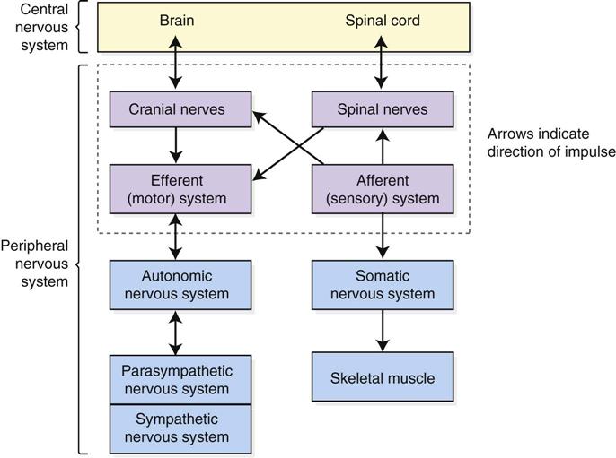
The nerve processes that connect the CNS with the various glands, muscles, and receptors in the body make up the peripheral nervous system. Functionally, the peripheral nervous system is divided into afferent and efferent portions. The afferent portion is composed of nerve cells that carry information from receptors in the periphery of the body to the CNS. The efferent system consists of nerve cells that carry impulses from the CNS to muscles and glands. Anatomically, the peripheral nervous system is composed of cranial nerves and spinal nerves.
The peripheral nervous system is also subdivided into a somatic nervous system and an autonomic nervous system (ANS). The somatic nervous system consists of efferent nerves that carry impulses from the CNS to skeletal muscle tissue. It is under conscious control and is therefore called voluntary. The ANS consists of efferent nerve cells that carry information from the CNS to cardiac muscle, glands, and smooth muscle. It is under unconscious control and is called involuntary. The ANS has two subdivisions, the sympathetic nervous system and the parasympathetic nervous system. Most tissues innervated by the ANS receive both sympathetic and parasympathetic fibers. In general, one division stimulates an activity by a receptor and the other inhibits the activity to serve as a method of checks and balances.
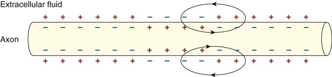
The fundamental unit of all branches and divisions of the nervous system is the neuron (nerve cell). Neurons have the amazing ability to transmit information from point to point. The second point may be nearby or at a great distance. Similar to all cells in the body, neurons have a nucleus surrounded by cytoplasm. Different from other cells, however, neurons have cellular extensions or processes called axons and dendrites. Axons carry electric-like messages away from the nerve cell, and dendrites carry electric-like messages toward the nerve cell (Figure 4-2). Transmission of these messages along nerve fibers occurs through a wave of charge reversal that moves down the fiber (Figure 4-3). The resting (polarized) fiber has positive charges lined up on the outside of its membrane and negative charges lined up on the inside of its membrane. When a stimulus of sufficient magnitude reaches the fiber, depolarization or charge reversal (positive in, negative out) occurs in a progressive wave down the fiber toward the synapse. Repolarization is the movement of charges back to their original positions.
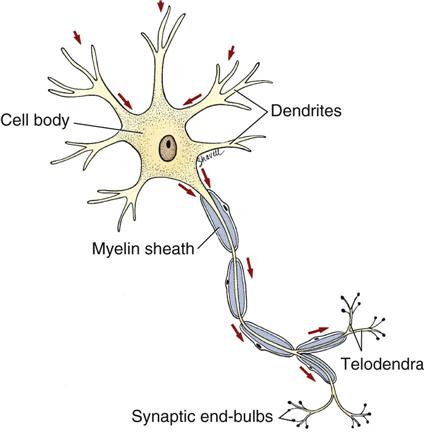
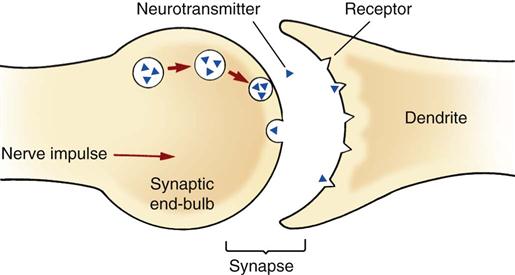
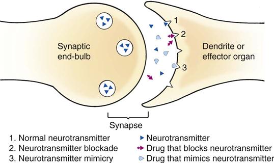
Axons may be short or long (up to 4 feet in humans), and they terminate, or end, in as many as 10,000 nerve endings called telodendra (Snyder, 1986). The large number of nerve endings allows for great variety in the number and type of connections made with other neurons. The synaptic end-bulbs of the telodendra pass nerve impulses to an adjacent structure (another neuron, gland, or muscle) by emitting a chemical messenger called a neurotransmitter into the gap or junction (synapse) between the nerve ending and the adjacent structure (Figure 4-4). Neurotransmitters then combine with receptors on the dendritic side of the synapse and cause a stimulatory or inhibitory effect. Dendrites may respond to neurotransmitters by generating a nerve impulse, which is conducted via the axon to the adjacent structure (neuron, gland, or muscle). Neurotransmitters can be mimicked or blocked by the use of appropriate drugs (Figure 4-5).
Nerve fibers (nerves) may have a large diameter (A fibers), a medium diameter (B fibers), or a small diameter (C fibers; Boothe, 2012). Fibers with large diameters conduct nerve impulses faster than those with small diameters. Fibers that are surrounded by the insulating substance called myelin also transmit impulses faster than nonmyelinated fibers. Type A and B fibers are generally myelinated fibers.
The most basic impulse conduction system through the nervous system is the reflex arc (Figure 4-6). The reflex arc is composed of the following:
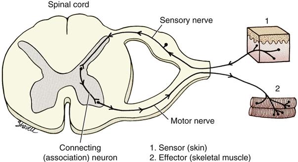
The receptor of the reflex arc may be located in a peripheral site—such as the skin—or in a central area—such as a muscle, tendon, or visceral organ. The sensory neuron carries the impulse from the receptor to the CNS. In the CNS, the sensory neuron synapses with interneurons in the spinal cord. These interneurons send the impulse to the brain for interpretation or send the impulse to a motor neuron. The motor neuron carries the message to an effector organ. If the impulse travels around the arc without going to the brain for analysis, the sequence of events is called a spinal reflex (see Figure 4-6). A spinal reflex can occur even if the spinal cord is completely severed. For example, a hemostat applied to the toe of a dog with a severed cord can cause the dog to withdraw its leg by means of the spinal reflex.
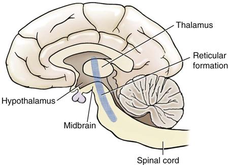
Areas of the brain that have importance to an understanding of the pharmacology of the CNS are illustrated in Figure 4-7. The cerebrum is responsible for higher functions of the brain, such as learning, memory, and interpretation of sensory input (e.g., vision and pain recognition). The thalamus serves as a relay center for sensory impulses from the spinal cord, brainstem, and cerebellum to the cerebrum. The thalamus also may be involved in pain interpretation. The hypothalamus serves as the primary mediator between the nervous system and the endocrine system through its control of the pituitary gland. The hypothalamus also controls and regulates the ANS. The medulla carries both sensory and motor impulses between the spinal cord and the brain. It contains centers that control vital physiologic activities, such as breathing, heartbeat, blood pressure, vomiting, swallowing, coughing, body temperature, hunger, and thirst. The reticular formation is a network of nerve cells scattered through bundles of fibers that begin in the medulla and extend upward through the brainstem. The reticular activating system is a part of the reticular formation, which functions to arouse the cerebral cortex and is responsible for consciousness, sleep, and wakefulness (DeLahunta, 1983).
In summary, nerve activity is usually described as the generation of nerve impulses that occurs in a dendrite or cell body and then travels down an axon by electric-like activity, which is similar to the passage of an electric current down a wire. When this current reaches a synapse, a chemical “bridge” or neurotransmitter allows the message to be passed to one or as many as thousands of other neurons. Neurotransmitter substances include acetylcholine, norepinephrine, dopamine, serotonin, and gamma-aminobutyric acid (GABA). These other neurons then carry the message to an interpretation center or a structure that takes appropriate action. CNS drugs act by mimicking or blocking the effects of neurotransmitters.
Autonomic Nervous System
The ANS is that portion of the nervous system that controls unconscious body activities. ANS fibers innervate smooth muscle, heart muscle, salivary glands, and other viscera. This system operates automatically and involuntarily to control visceral functions, such as gastrointestinal (GI) motility, rate and force of the heartbeat, secretion by glands, sizes of the pupils, and various other involuntary functions and characteristics. In contrast to the somatic nervous system, the ANS has two subdivisions: parasympathetic (cholinergic) and sympathetic (adrenergic). The sympathetic division regulates energy-expending activities (fight-or-flight responses), and the parasympathetic division regulates energy-conserving activities.
The ANS has two neurons that carry impulses to target structures (in contrast to the somatic nervous system, which has only one). The cell body of the first neuron arises in the CNS—in the thoracolumbar cord for the sympathetic nervous system and in the craniosacral cord for the parasympathetic nervous system (Figure 4-8). The axon of the first neuron leaves the CNS and travels to a ganglion, where it synapses with dendrites of the second neuron. This second neuron then travels to the target structure (Figure 4-9). Axons of the first neuron are called preganglionic, and those of the second are called postganglionic. The synapse between the preganglionic neuron and the postganglionic neuron is called the ganglionic synapse.
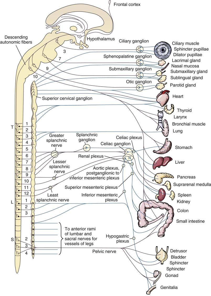
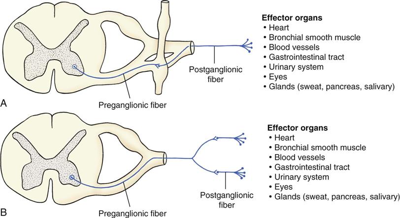
Preganglionic fibers of the sympathetic nervous system are short. They end in ganglia adjacent to the spinal cord. The only exception is the preganglionic fiber to the adrenal medulla. The adrenal medulla itself is analogous to a postganglionic fiber because it releases epinephrine and norepinephrine directly into the bloodstream when stimulated by preganglionic fibers. Postganglionic sympathetic fibers are long.
Preganglionic fibers of the parasympathetic nervous system are generally long. They travel to ganglia located in the wall of the target organ. Postganglionic fibers are consequently short.
Normally, target sites of the ANS have both sympathetic and parasympathetic innervation. The physiologic functions of the two systems usually oppose each other, thereby bringing about a state of balance. When this balance is disrupted, drug therapy may be indicated to restore the balance. The adrenal medulla, sweat glands, and hair follicles have only sympathetic fibers.
Stimulation of the sympathetic nervous system causes an increase in heart rate and respiratory rate, a decrease in GI activity, dilation of the pupils, constriction of blood vessels in smooth muscle, dilation of blood vessels in skeletal muscle, dilation of bronchioles, and an increase in blood glucose levels. These actions prepare an animal to fight or to flee. On the other hand, stimulation of the parasympathetic nervous system causes a decrease in heart rate and respiratory rate, an increase in GI activity, constriction of the pupils, and constriction of the bronchioles.
Receptors of the sympathetic (adrenergic) nervous system are subdivided as follows (Figure 4-10):
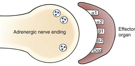
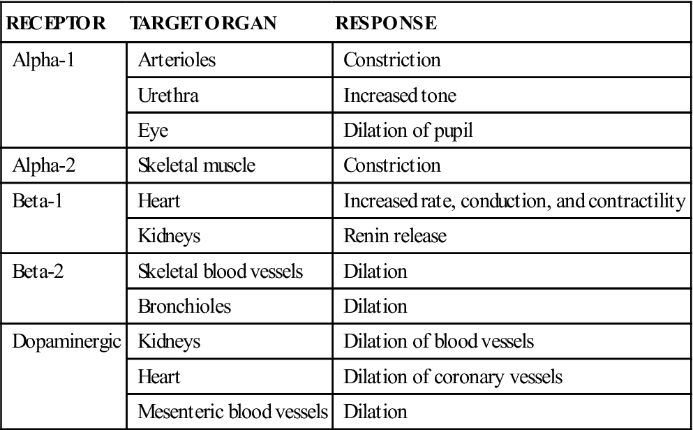
Generally, alpha receptors are stimulatory and beta receptors are inhibitory (Table 4-1). The parasympathetic (cholinergic) nervous system has nicotinic and muscarinic receptors. Effector organs have one or a combination of these receptors. A drug’s effect is determined by the number of receptors in the effector and the drug’s specificity for the receptor (Williams and Baer, 1990).
TABLE 4-1
| RECEPTOR | TARGET ORGAN | RESPONSE |
| Alpha-1 | Arterioles | Constriction |
| Urethra | Increased tone | |
| Eye | Dilation of pupil | |
| Alpha-2 | Skeletal muscle | Constriction |
| Beta-1 | Heart | Increased rate, conduction, and contractility |
| Kidneys | Renin release | |
| Beta-2 | Skeletal blood vessels | Dilation |
| Bronchioles | Dilation | |
| Dopaminergic | Kidneys | Dilation of blood vessels |
| Heart | Dilation of coronary vessels | |
| Mesenteric blood vessels | Dilation |
The primary neurotransmitters for adrenergic sites are norepinephrine, epinephrine, and dopamine. Epinephrine equally stimulates alpha and beta receptors and is therefore a potent stimulator of the heart and an equally powerful dilator of bronchioles. Acetylcholine is the neurotransmitter at sympathetic postganglionic fibers to sweat glands and the smooth muscle of blood vessels (muscarinic sites).
The neurotransmitter for cholinergic sites is acetylcholine. Acetylcholine combines with both nicotinic and muscarinic receptors.
Cholinergic sites are found in both the sympathetic and parasympathetic nervous systems. Nicotinic receptors are found in all autonomic ganglia, in the adrenal medulla, and at the neuromuscular junction of the somatic nervous system. Muscarinic receptors are found at the synapse of postganglionic fibers of the parasympathetic nervous system and at a few of the sympathetic postganglionic fibers.
How Drugs Affect the Autonomic Nervous System
Autonomic drugs bring about their effects by influencing the sequence of events that involve neurotransmitters. Most autonomic drugs bring about this alteration of events by doing the following:
Classes of Autonomic Nervous System Agents
 Cholinergic Agents
Cholinergic agents are drugs that stimulate receptor sites mediated by acetylcholine. They achieve these effects by mimicking the action of acetylcholine (direct-acting) or by inhibiting its breakdown (indirect-acting). Cholinergic agents are also called parasympathomimetic because their effects resemble those produced by stimulating parasympathetic nerves.
Clinical Uses
Cholinergic agents do the following:
Direct-Acting Cholinergics
Indirect-Acting Cholinergic (Anticholinesterase) Agents
• Edrophonium (Tensilon). Edrophonium is used to diagnose myasthenia gravis.
• Physostigmine (Antilirium, Eserine). Uses of this product are similar to those of neostigmine.
• Demecarium (Humorsol). This drug is used in the preventive management of glaucoma.
• Pyridostigmine (Mestinon). This drug is used for the treatment of myasthenia gravis.
Adverse Side Effects
Adverse side effects of the cholinergic drugs may include bradycardia, hypotension, heart block, lacrimation, diarrhea, vomiting, increased intestinal activity, intestinal rupture, and increased bronchial secretions.
 Cholinergic Blocking Agents (Anticholinergic)
Cholinergic blocking agents are drugs that block the action of acetylcholine at muscarinic receptors of the parasympathetic nervous system.
Clinical Uses
Clinical uses of these drugs include the following:
• Treatment of diarrhea and vomiting via a decrease in GI motility
• Drying of secretions and prevention of bradycardia before anesthesia
• Dilation of the pupils for ophthalmic examination
• Relief of ciliary spasm of the eye
The belladonna alkaloids of the deadly nightshade family of plants have been used as drugs for centuries and represent the prototype for this category of agents.
Dosage Forms
Adverse Side Effects
Adverse side effects of the cholinergic blockers are dose related. Overdoses can cause drowsiness, disorientation, tachycardia, photophobia, constipation, anxiety, and burning at the injection site.
 Technician Notes
 Adrenergic (Sympathomimetic) Agents
Adrenergic (sympathomimetic) agents bring about action at receptors mediated by epinephrine or norepinephrine. Adrenergic agents may be classified as catecholamines or noncatecholamines, and either category can also be classified according to the specific receptor types activated (alpha-1, alpha-2, beta-1, beta-2). In most cases, alpha receptor activity causes an excitatory response (except in the GI tract), and beta stimulation causes an inhibitory response (except in the heart). Adrenergic activity is a complex subject, and more advanced texts should be consulted for a thorough explanation.
Clinical Uses
Adrenergic agents are used for the following purposes:
• To stimulate the heart to beat during cardiac arrest
• To reverse the hypotension and bronchoconstriction of anaphylactic shock
• To strengthen the heart during congestive heart failure
• To correct hypotension through vasoconstriction
• To reduce capillary bleeding through vasoconstriction
• To treat urinary incontinence
• To reduce mucous membrane congestion (vasoconstriction) in allergic conditions
Dosage Forms
Adverse Side Effects
These may include tachycardia, hypertension, nervousness, and cardiac arrhythmias. Hypertension, arrhythmia, and pulmonary edema may occur with an overdose.
 Technician Notes
Epinephrine is normally packaged as a 1 : 1000 dilution. Many clinicians prefer a 1 : 10,000 dilution for treating cardiac arrest. Mix 1 mL of the original dilution with 9 mL of sterile water to prepare a 1 : 10,000 dilution. Epinephrine is stored under refrigeration. A note should be placed in the emergency crash cart specifying the location of this drug.
 Adrenergic Blocking Agents
Adrenergic blocking agents are used to disrupt the activity of the sympathetic nervous system. They are classified according to the site of their action as an alpha blocker, beta blocker, or ganglionic blocker. Drugs usually block only one category of receptor.
Alpha Blockers
Alpha blockers have had limited use in veterinary medicine. Phenoxybenzamine has been advocated by some clinicians for the treatment of laminitis in horses and urethral obstruction in cats. Yohimbine is used for xylazine antagonism.
Clinical Uses
See section, Dosage Forms.
Dosage Forms
• Phenoxybenzamine (Dibenyline). Phenoxybenzamine is a hypotensive (vasodilator) agent.
• Aacepromazine. This tranquilizer acts as an alpha blocker and causes vasodilation.
• Prazosin (Minipress). Prazosin is a hypotensive agent.
• Yohimbine (Yobine). Yohimbine is used as an antidote for xylazine toxicity.
• Atipamezole (Antisedan). Atipamezole is a reversal agent for dexmedetomidine.
Adverse Side Effects
Adverse side effects may include hypotension (phenoxybenzamine, tranquilizers, prazosin), tachycardia (phenoxybenzamine), muscle tremors (yohimbine), and seizures (acepromazine).
Beta Blockers
Beta blockers are used to treat glaucoma, arrhythmias, and hypertrophic cardiomyopathy.
Clinical Uses
See section, Dosage Forms.
Dosage Forms
• Timolol (Timoptic). Timolol is an ophthalmic preparation used to treat glaucoma.
• Atenolol (Tenormin). Used in a similar way to propranolol.
• Carteolol (Ocupress). Carteolol is a human labeled antiglaucoma medication.
• Levobunolol (Betagan). Levobunolol is a human labeled antiglaucoma medication.
• Metipranolol (OptiPranolol). Similar to the previous two products.
Adverse Side Effects
These include bradycardia, hypotension, worsening of heart failure, bronchoconstriction, heart block, and syncope.
Ganglionic Blockers
Ganglionic blockers are seldom used in veterinary medicine.
Central Nervous System
CNS drugs have various uses in veterinary medicine. Depressant drugs are used to tranquilize or sedate animals to facilitate restraint or anesthetic procedures. They are also used to control pain, to induce anesthesia, and to prevent or control seizures. CNS drugs are also available to antagonize (reverse) the effects of some depressant drugs. Another group of CNS agents is used to stimulate the CNS to treat cardiac or respiratory depression or arrest. Euthanasia drugs allow veterinarians to provide a quick and painless end to hopeless medical situations.
Drugs that affect the CNS generally cause depression or stimulation. They are thought to generate these changes by altering nerve impulse transmissions between the spinal cord and the brain or within the brain itself. Altering impulse transmissions within the thalamus could prevent messages regarding painful stimuli from reaching interpretation centers within the cerebrum. Interfering with impulses within the reticular activating system could alter levels of consciousness or wakefulness (Ganong, 2003). The changes that occur in the transmission of nerve impulses as a result of administration of CNS drugs are probably brought about by altered neurotransmitter activity.
The categories of CNS drugs that are covered in this chapter include the following:
< div class='tao-gold-member'>
Stay updated, free articles. Join our Telegram channel

Full access? Get Clinical Tree


