Drugs Used in Hormonal, Endocrine, and Reproductive Disorders
Learning Objectives
After studying this chapter, you should be able to
1. Discuss the control mechanisms (physiology) of the endocrine system.
3. List the reasons why hormones are clinically used.
4. Describe the difference between an endogenous and an exogenous hormone.
5. Describe the location and functions of the pituitary gland.
6. Differentiate between a positive and a negative feedback control mechanism.
7. Describe a neurohormonal reflex.
9. Describe the uses and classes of drugs that affect uterine contractility.
10. Define pheromone and give an example.
11. Describe the location, function, and hormonal products of the thyroid gland.
12. Describe the hormonal treatment of hypothyroidism and hyperthyroidism.
13. List the two forms of spontaneous Cushing’s syndrome.
14. List two or more drugs used to treat Cushing’s syndrome.
15. List the endogenous source of insulin and its metabolic effects.
16. List the classes of insulin products and their general characteristics.
17. Describe the method of action of the growth promoters.
Key Terms
Anabolism
Analogue
Cushing’s syndrome
Dystocia
Endometrium
Euthyroid
Feed Efficiency
Feedback
Gonadotropin
Hypophyseal Portal System
Iatrogenic
Involution
Levo Isomer
Myofibril
Nitrogen Balance
Primary Hypothyroidism
Releasing Factor (Releasing Hormone)
Trophic Hormone
Introduction
The traditional definition of the endocrine system states that it is composed of organs (glands) or groups of cells that secrete regulatory substances (hormones) directly into the bloodstream. This definition has now been extended to include regulatory substances that are distributed by diffusion across cell membranes.
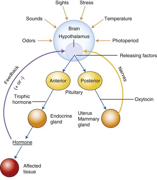
The endocrine system and the nervous system constitute the two major control mechanisms of the body. These two control mechanisms are linked together through the complex integrating action of the hypothalamus (Figure 9-1). Coordination of these two systems allows an individual to adapt its reproductive and survival strategies to changes in the environment.
Endocrine glands include the pituitary, adrenals, thyroid, ovaries, testicles, pancreas, and kidneys. These glands produce hormones that are carried to target organs, where they influence the physiologic activity of these structures.
Hormones generally are administered to animals for one of two reasons: (1) to correct a deficiency of that hormone or (2) to obtain a desired effect (e.g., to postpone estrus). Hormones that are administered to an animal are called exogenous hormones, whereas those produced naturally in the body are endogenous hormones.
Anatomy and Physiology
Pituitary Gland
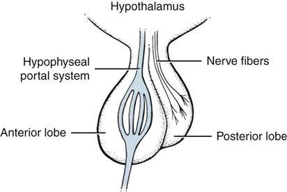
The pituitary gland has been called the master gland of the endocrine system because of the control it exerts over the regulation of this system. It is located at the base of the brain just ventral to the hypothalamus and is connected to the brain by a stalk. It is divided into two main lobes—an anterior lobe (adenohypophysis), which arises from the embryologic pharynx, and a posterior lobe (neurohypophysis), which arises from the brain (Figure 9-2).
The hypothalamus exerts control over the anterior pituitary through the transport of releasing hormones, or factors, down the hypophyseal portal system. In the anterior pituitary, these releasing factors cause the secretion of trophic hormones into the circulation. Trophic hormones produced by the anterior pituitary include thyroid-stimulating hormone (TSH), adrenocorticotropic hormone (ACTH), luteinizing hormone (LH), follicle-stimulating hormone (FSH), prolactin (LTH), and growth hormone (GH or somatotropin). These trophic hormones are sometimes called indirect-acting hormones because they cause their target organ to produce a second hormone, which in turn influences a second target organ or tissue (Table 9-1). For example, TSH stimulates the thyroid gland to produce triiodothyronine (T3) and tetraiodothyronine (T4), which are hormones that in turn influence the metabolic rate of all tissues in the body.
TABLE 9-1
| SOURCE AND NAME | TARGET AND ACTIONS |
| Anterior Lobe | |
| Thyroid-stimulating hormone (TSH) | Stimulates the thyroid to produce T3/T4 |
| Follicle-stimulating hormone (FSH) | Stimulates ovarian follicle growth (female) and spermatogenesis (male) |
| Luteinizing hormone (LH) | Stimulates ovulation (female) and testosterone production (male) |
| Growth hormone (somatotropin) | Accelerates body growth and increases milk production |
| Adrenocorticotropic hormone (ACTH) | Stimulates production of corticosteroids by adrenal cortex |
| Posterior Lobe | |
| Oxytocin | Stimulates uterine contraction and milk letdown |
| Vasopressin (antidiuretic hormone, ADH) | Stimulates water retention |
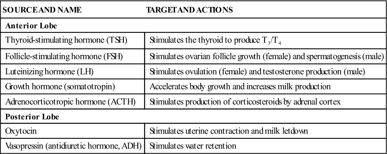
The two hormones of the posterior pituitary are vasopressin (antidiuretic hormone) and oxytocin. These hormones are produced in the hypothalamus and subsequently travel down nerve fibers to the posterior pituitary, where they are stored for release into the circulation. The hormones of the posterior pituitary are called direct-acting hormones because they produce the desired activity (e.g., contraction of the uterus) directly in the target organ.
Control of the Endocrine System
Feedback Mechanism
The nervous system is sensitive to levels of hormones through a mechanism called feedback. By this mechanism, the plasma level of a particular hormone controls the activity of the gland that produces it. The feedback may be negative or positive (Figure 9-3).
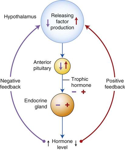
With negative feedback, high plasma levels of a hormone are sensed by the hypothalamus, which then reduces the amount of the appropriate releasing factor (or hormone). A decreased amount of releasing factor reduces the amount of trophic hormone released from the pituitary, causing less activity in the organ that is producing the hormone in question. The overall effect is to lower the amount of the hormone in the plasma.
In the positive feedback scheme, low levels of a hormone are sensed by the hypothalamus, and release of the appropriate releasing factor increases. Increased amounts of the corresponding trophic hormone are then secreted, causing increased activity in the target organ and a corresponding rise in the plasma levels of the hormone.
Neurohormonal Reflex
The neurohormonal reflex applies to the release of oxytocin by the posterior pituitary. The first step in this reflex can be initiated by (1) stimulation of the udder by a nursing calf or by preparation of the udder for milking, (2) stimulation of the uterus and vagina in parturition, or (3) stimulation of the cerebral cortex by sensory stimuli associated with nursing or milking.
Control of the Reproductive System
The reproductive (estrus) cycle in animals traditionally has been divided into four stages called proestrus, estrus, diestrus, and anestrus. The cycle also may be divided into a follicular phase and a luteal phase. In the follicular phase, the cycle is under the influence of estrogen produced by a developing follicle, and in the luteal phase, it is under the influence of progesterone made by the corpus luteum.
Control of the reproductive system is coordinated in the hypothalamus, where the gonadotropin-releasing hormone (GnRH) is produced in response to various stimuli (Figure 9-4). These stimuli can include day–night length (photoperiod), pheromones, and positive and negative internal feedback mechanisms. GnRH causes the release of FSH and LH from the anterior pituitary.
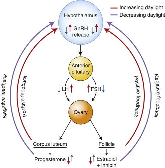
FSH causes the growth and maturation of a follicle, which begins to produce increasing amounts of estrogen as it matures. Estrogen causes the changes that occur in proestrus and estrus, including the behavioral characteristics associated with estrus (e.g., standing to be mounted). The follicle also produces inhibin, which—along with estrogen—serves as negative feedback to the hypothalamus to inhibit the release of GnRH.
LH release causes ovulation of the mature follicle and the formation of a corpus luteum in its place. This event signals the beginning of diestrus and the beginning of the luteal phase of the cycle. The corpus luteum produces progesterone, which prepares the uterus for pregnancy. Once pregnancy occurs, the corpus luteum maintains a uterine environment conducive to normal progression of the pregnancy. Progesterone levels in the blood serve as negative feedback to prevent the release of GnRH and the development of new follicles during pregnancy.
When the gestation period nears its end, the fetus begins to produce increasing amounts of ACTH. ACTH causes increased amounts of cortisol to be produced by the adrenal glands. The increased cortisol levels result in increased production of estrogen and prostaglandin by the uterus. These two substances sensitize the uterus to the contraction-producing effects of oxytocin and allow parturition to begin. Prostaglandin also causes the breakdown (lysis) of the corpus luteum at the end of pregnancy and at the end of diestrus if pregnancy does not occur.
Hormonal Drugs Associated with Reproduction
 Gonadotropins and Gonadal Hormones
Products in this category are used in veterinary medicine for various reasons. Some of these include synchronization of estrus, suppression of estrus, induction of estrus, treatment of cystic ovaries, and termination of pregnancy.
Gonadotropins
Gonadotropins are drugs that act similarly to GnRH, LH, or FSH. Gonadotropins cause the release of LH and FSH or cause activity similar to that of LH or FSH. LH may be prepared from the pituitary glands of slaughtered animals, from the urine of pregnant women in the form of human chorionic gonadotropin (hCG), or in pure form through recombinant techniques. FSH may be obtained from pituitary glands (FSH-P), from the serum of pregnant mares (PMS) between the 40th and 140th days of pregnancy, or from recombinant sources. GnRH is prepared synthetically.
FSH that is released endogenously by the anterior pituitary causes growth and maturation of the ovarian follicle in females and spermatogenesis in males. LH, also released by the anterior pituitary, causes ovulation in females and production of testosterone in males.
Gonadorelin
Gonadorelin (GnRH) is produced endogenously by the hypothalamus. Gonadorelin causes the release of FSH and LH by the anterior pituitary.
Clinical Uses
Gonadorelin is used to treat cystic (follicular) ovaries in dairy cattle. It has also been used in cats and horses (with limited success) to induce estrus.
Dosage Forms
Adverse Side Effects
These are minimal with the use of this product.
Chorionic Gonadotropin
hCG is a hormone secreted by the uterus and obtained from the urine of pregnant women. It mimics the effects of LH, although it has limited FSH activity. In males, it stimulates the production of male hormones by the testicles and may facilitate descent of the testicles.
Clinical Uses
Chorionic gonadotropin is used to treat cystic ovaries (nymphomania) in dairy cattle. In males, it has been used to treat cryptorchidism and infertility caused by low testosterone levels.
Dosage Forms
Adverse Side Effects
These are limited but may include hypersensitivity reaction and abortion in mares if given before the 35th day of pregnancy.
Follicle-Stimulating Hormone
FSH causes growth and maturation of the ovarian follicle. FSH-P, prepared from the pituitary glands from slaughtered animals, is no longer available. Purified recombinant human FSH may be obtained, but it is expensive. Chorionic gonadotropin is readily available and causes FSH-like activity (and LH activity) and is more likely to be used in reproductive cases.
Clinical Uses
FSH has been used in veterinary medicine to induce superovulation and for out-of-season breeding.
Dosage Form
Adverse Side Effects
These include endometrial hyperplasia, superovulation, and follicular cysts.
Estrogens
Estrogens are a group of hormones synthesized by the ovaries and—to a lesser extent—by the testicles, adrenal cortex, and placenta. Estrogens are classified as sex steroids and are synthesized from a cholesterol precursor. Estrogens are necessary for normal growth and development of the female gonads. They cause secondary female characteristics and are responsible for female sex drive. These hormones inhibit ovulation, increase uterine tone, and cause proliferation of the endometrium.
Clinical Uses
In cattle, estrogens are used to treat persistent corpus luteum, to expel purulent material from the uterus, to expel retained placentas and mummified fetuses, and to promote weight gain. In dogs, estrogens may be used to control urinary incontinence. In horses, they may be used for induction of estrus in the nonbreeding season.
Dosage Forms
• Estradiol cypionate (ECP) injection
• Estradiol cypionate for injection (Depo-Estradiol), human label
• Estradiol valerate for injection (Delestrogen), human label
• Diethylstilbestrol (DES) compounded capsules and tablets
• Estriol (Incurin), for control of urinary incontinence in spayed dogs
• Implants to promote weight gain (discussed in a later section)
Adverse Side Effects
These include severe anemia, prolonged estrus, genital irritation, and follicular cysts.
 Technician Notes
Androgens
Androgens are male sex hormones produced in the testicles, the ovaries, and the adrenal cortex. Similar to the other gonadal hormones, they have a steroidal parent molecule. These hormones are necessary for growth and development of the male sex organs. They cause secondary male sex characteristics and produce male libido. The androgens promote tissue anabolism, weight gain, and red blood cell formation.
Methyltestosterone, Testosterone Cypionate, Testosterone Enanthate, and Testosterone Propionate
These injectable testosterone products are available under a human label.
Clinical Uses
These androgens are used to treat urinary incontinence in male dogs and to increase libido and fertility in domestic animals (with generally poor results).
Dosage Forms
Adverse Side Effects
These are uncommon when the products are used as directed.
 Technician Notes
Testosterone products are now Class III controlled substances.
Mibolerone
Mibolerone is an androgen used for prevention of estrus in dogs. Mibolerone blocks the release of LH by the pituitary and prevents complete development of the follicle. Ovulation does not occur.
Clinical Uses
This product is used for prevention of estrus in adult female dogs and for treatment of pseudocyesis.
Dosage Forms
Adverse Side Effects
Adverse side effects reported in the product insert include premature epiphyseal closure and vaginitis in immature females. In mature females, vulvovaginitis, clitoral hypertrophy, riding behavior, increased body odor, and various other side effects have been reported. It is further reported that side effects usually resolve with discontinuation of therapy.
 Technician Notes
Mibolerone should not be used in cats because of a very low margin of safety in this species.
Progestins
Progestins are a group of compounds that are similar in effect to progesterone. Endogenous progestins are produced by the corpus luteum. They cause increased secretions by the endometrium, decreased motility in the uterus, and increased secretory development in the mammary glands. They also inhibit the release of gonadotropins by the pituitary to produce an inactive ovary. In some situations, they can cause elevated blood glucose levels (antiinsulin effect) or serious suppression of the adrenal glands. These hormones are used clinically to suppress estrus and to treat false pregnancy, behavioral disorders, and progestin-responsive dermatitis. The root “gest” often allows name recognition of the progestins.
Megestrol Acetate
Megestrol acetate is a synthetic progestin labeled for use in dogs. It is used, however, in cats for some behavioral and dermatologic conditions.
Clinical Uses
Megestrol acetate is labeled for use in dogs to control estrus, treat false pregnancy, prevent vaginal hyperplasia, treat severe galactorrhea, and control unacceptable male behavior. Megestrol acetate has been used in cats for various dermatologic and behavioral problems and for suppression of estrus.
Dosage Forms
Adverse Side Effects
These can include hyperglycemia, adrenal suppression, endometrial hyperplasia, and increased appetite.
 Technician Notes
Clients should be made aware of the potential dangers associated with the use of megestrol acetate and should be asked to report any changes in their pet’s health status that occur after initiation of therapy.
Medroxyprogesterone Acetate
Medroxyprogesterone acetate (MPA) is a human label progestin that has been used to treat certain behavioral and dermatologic problems and to suppress estrus in dogs and cats.
Clinical Uses
MPA is used for (1) treatment of behavioral problems, such as aggression, roaming, spraying, or mounting in males, and (2) treatment of certain dermatologic conditions.
Dosage Forms
Adverse Side Effects
These are potentially numerous and include pyometra, personality changes, depression, lethargy, mammary changes, and increased appetite.
 Technician Notes
Progestins should be administered with strict adherence to an accepted protocol to minimize side effects such as pyometra.
Altrenogest
Altrenogest is an oral progestin labeled for use in horses and swine. This drug is used to suppress estrus in mares and sexually mature gilts. Mares stop cycling within 3 days of treatment and begin cycling again 4 to 5 days after treatment is stopped. It is also used to manage other reproductive conditions that are listed later.
Clinical Uses
Altrenogest is used to suppress estrus for synchronization, to suppress estrus for long periods, or to maintain pregnancy in mares with low levels of progesterone.
Dosage Form
Adverse Side Effects
These have been reported as minimal when altrenogest is used correctly.
 Technician Notes
Altrenogest can be absorbed through the skin and should be used with great caution by pregnant women or anyone with vascular disorders. Read the label carefully before using.
Norgestomet
Norgestomet is a synthetic progestin that is used in combination with an estrogen (estradiol valerate) for synchronization of estrus in beef cows and nonlactating dairy cows. A treatment consists of one implant and an injection at the time of implantation.
Clinical Uses
Norgestomet is used for synchronization of estrus in cattle.
Dosage Form
< div class='tao-gold-member'>
Stay updated, free articles. Join our Telegram channel

Full access? Get Clinical Tree


