Drugs Used in Gastrointestinal System Disorders
Learning Objectives
After studying this chapter, you should be able to
1. Exhibit a basic understanding of the anatomy and physiology of the gastrointestinal (GI) system.
2. Describe the various mechanisms of control of the GI system.
3. Explain the difference between vomiting and diarrhea.
4. Exhibit a working knowledge of drugs that induce vomiting and those that inhibit it.
5. List and describe antiulcer medications used in veterinary medicine.
6. Explain the pathophysiology of diarrhea and list the medications used to control this condition.
7. List the different categories of laxatives and explain their respective mechanisms of action.
8. List the two basic categories of GI prokinetics and stimulants.
9. Explain why digestive enzymes are used.
10. Discuss the use of antibiotics and antiinflammatory agents in GI disease.
11. List the categories of oral products and give an example from each category.
Key Terms
Adsorbent
Anticholinergic
Chemoreceptor trigger zone
Cholinergic
Dentifrice
Emesis
Hematemesis
Melena
Motilin
Parietal cell
Peristalsis
Regurgitation
Segmentation
Vomiting center
Introduction
Problems of the gastrointestinal (GI) system are common reasons for visits to a veterinary practice. These problems include regurgitation, vomiting, diarrhea, weight loss, colic, bloating, flatulence, abnormal stools, and constipation. Because veterinary technicians are expected to answer clients’ questions about the GI tract, administer therapeutic GI medications, and monitor the response to GI medications, they must be knowledgeable about this system. They should have a basic knowledge of GI anatomy, physiology, pathophysiology, therapeutic principles, and medications.
Anatomy and Physiology
Anatomic and physiologic differences between the GI systems of different animal species are greater than for any other organ system. Despite these differences, the functions are basically the same in each species: (1) intake of food and fluid into the body, (2) absorption of nutrients and fluid, and (3) excretion of waste products. A discussion of the anatomy and physiology of the GI tract with an emphasis on similarities and differences between species follows.
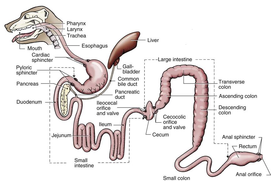
The basic structures of the GI tract include (depending on the species) the mouth, teeth, tongue, salivary glands, esophagus, outpocketings of the esophagus (i.e., crop, reticulum, rumen, and omasum), stomach, liver, pancreas, duodenum, jejunum, ileum, cecum, colon, rectum, and anus.
Carnivorous or omnivorous species (e.g., cats, dogs, and primates) often are described as monogastric or simple-stomach animals because they have no outpocketings or forestomachs arising from the basic configuration (Figure 8-1). The function of the stomach in these monogastric animals is primarily to store ingested material and to begin some enzymatic breakdown of protein. The salivary glands begin enzymatic digestion by producing enzymes that break down starch into simpler carbohydrates. Pancreatic enzymes delivered to the duodenum break down fats, carbohydrates, and proteins, and sodium bicarbonate from the pancreas neutralizes hydrochloric acid from the stomach. Bile salts, produced in the liver and delivered to the duodenum, aid in digestion by emulsifying fats. Bile is stored in the gallbladder, which is absent in some animals (e.g., horses and rats). Digestion and its control mechanisms are complex, and students should consult an appropriate text for further information.
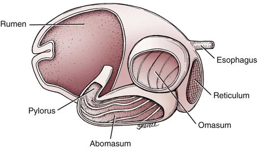
Ruminant animals are herbivorous and have a GI system characterized by three forestomachs, the reticulum, rumen, omasum, and a “true” stomach—the abomasum (Figure 8-2). The reticulum receives ingested material and passes it to the rumen, where it is mixed and acted on by microorganisms to digest cellulose and other coarse plant material (roughage). Some refer to the rumen as a “fermentation vat,” where microorganisms break down coarse feeds into forms that can be used by the simple stomach portion of the GI system in ruminants. Partially digested material (cud) in the rumen is regurgitated and remasticated to further facilitate digestion. In an immature ruminant, an esophageal groove allows milk to bypass the rumen and flow directly into the abomasum, and the rumen gains full function only after several months.
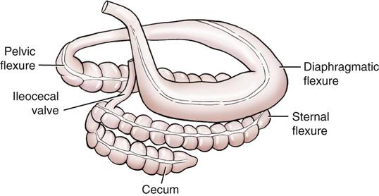
Equines, rabbits, and some rodents are chiefly herbivorous animals that have a monogastric GI configuration. They possess, however, a large cecum, which is capable of limited roughage digestion (hindgut fermentation) (Figure 8-3).
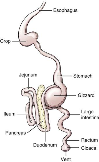
Birds have an outpocketing of the esophagus called the crop, which is used for food storage. They also have a ventriculus, or gizzard, which serves to grind coarse food material (Figure 8-4).
The small intestine comprises three sections: the duodenum, which has a sharp bend and in which the pancreas is located; the long and highly coiled jejunum; and the short ileum, which connects to the large intestine. In the small intestine, contents passing from the stomach are mixed with intestinal secretions, pancreatic juice, and bile. The digestive process that began in the mouth and stomach is completed in the small intestine. Products of this process are absorbed together with most of the vitamins and a great deal of fluid. Villi and microvilli protrude from the mucosal surface into the lumen of the small intestine and greatly enhance the absorptive process.
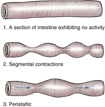
Movements of the small intestine mix the intestinal contents, called chyme, and move them toward the large intestine. Normal intestinal motility includes two different patterns—peristalsis and segmentation (Figure 8-5). Peristalsis is a wave of contractions that propels contents along the digestive tract. Segmentation is a periodic, repeating pattern of intestinal constrictions that serves to mix and churn the contents.
The colon has a considerably larger diameter than the small intestine. The colon is connected to the ileum and the cecum through the ileocecocolic valve. The surface of the colon may exhibit one or more longitudinal bands (depending on the species) called teniae. The wall of the colon may also form outpocketings, called haustra. The colon of monogastric animals has an ascending portion, a transverse portion, and a descending portion that leads into the rectum. Functions of the colon include absorption of water, synthesis of certain vitamins, and storage of waste material.
Movements of the colon include peristalsis and segmentation (as in the small intestine), as well as a third type called mass action contraction (Ganong, 2003). Mass action contraction is a result of simultaneous contraction of smooth muscle over a large area and serves to move fecal material from one portion of the colon to another and from the colon into the rectum.
Regulation of the Gastrointestinal System
Regulation of GI system activity is complex but can be said to be under the influence of the following three basic control systems:
1. The autonomic nervous system (ANS).
• Stimulation of various intrinsic receptors in the GI tract, such as the myenteric plexus (stretch receptor), also may increase peristaltic activity. Some physiologists consider the intrinsic receptors (myenteric plexus and Meissner’s plexus) to be a third portion of the ANS called the enteric nervous system (Ganong, 2003).
Another factor that can have a major influence on GI activity is the presence of bacterial endotoxins. Endotoxins are components of the bacterial cell wall of certain bacteria (often gram-negative bacteria) that may increase the permeability of intestinal blood vessels and cause increased fluid loss and fever.
Vomiting
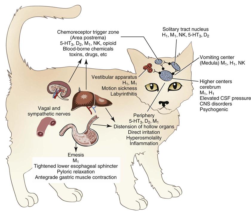
Vomiting is forceful ejection of the contents of the stomach, and sometimes the contents of the proximal small intestine, through the mouth. Vomiting is initiated by activation of the vomiting (emetic) center in the medulla of the brain. The vomiting center is connected by nerve pathways to the chemoreceptor trigger zone (CRTZ), the cerebral cortex, and peripheral receptors in the pharynx, GI tract, urinary system, and heart. Impulses from any of these areas activate the vomiting reflex; this requires a coordinated effort of the GI, musculoskeletal, respiratory, and nervous systems. Impulses may be generated by (1) pain, excitement, or fear (cortex); (2) disturbances of the inner ear (CRTZ); (3) drugs such as apomorphine and digoxin (CRTZ); (4) metabolic conditions such as uremia, ketonemia, or endotoxemia (CRTZ); and (5) irritation of peripheral receptors.
Multiple neurotransmitters are involved in the vomiting reflexes. Some of those include histamine (H1), dopaminergic (D2), serotonergic (5-HT3), neurokinin (NK), acetylcholine (muscarinic, M1), substance P, and other neurotransmitters. Agents that prevent emesis (antiemetics) exert their effects by blocking one or more of these neurotransmitters (Figure 8-6).
Occasional vomiting by a dog or cat is considered normal. However, persistent vomiting is not normal. Horses and rats do not normally vomit. Persistent vomiting can cause serious problems such as resultant dehydration, electrolyte disturbances, and acid–base imbalances. Sizable quantities of sodium, potassium, and chloride are lost in vomit. However, potassium loss is usually the most significant abnormality.
 Emetics
Emetics are drugs that induce vomiting. Emetics are administered to animals that have ingested toxins, but they must be used carefully to avoid serious complications. Emetics must be administered within 2 to 4 hours of the toxic ingestion to be effective. Emetics should not be used in animals that (1) are comatose or are having a seizure; (2) have depressed pharyngeal reflexes; (3) are in shock or have dyspnea; or (4) have ingested strong acid, alkali, or other caustic substances. Obviously, emetics should not be given to animals that do not normally vomit, such as rabbits, some rodents, and horses. Emetics usually remove about 80% of the stomach contents. Therefore the animal should be closely monitored for signs of toxicity after induced vomiting (Plumb, 2011).
Emetics are classified according to their site of action. Those acting on the CRTZ are categorized as centrally acting, and those that act on peripheral receptors are locally acting.
Centrally Acting Emetics
Apomorphine
Apomorphine is a morphine derivative that stimulates dopamine receptors in the CRTZ, which then activates the vomiting center. This drug is poorly absorbed after oral administration and is therefore usually administered topically in the conjunctival sac or parenterally. Vomiting follows rapidly after intravenous administration, 5 to 10 minutes after intramuscular injection, and variably (10 to 20 minutes) after conjunctival administration.
Clinical Uses
Apomorphine is used primarily for induction of vomiting in dogs. It is considered by many to be the emetic of choice for dogs. Its use in cats is controversial and possibly is contraindicated. Xylazine, which is safer than apomorphine, is effective as an emetic in most cats.
Dosage Forms
Apomorphine is a Class II controlled substance.
Adverse Side Effects
These include protracted vomiting, restlessness, and depression.
 Technician Notes
Xylazine
Although xylazine is not classified as an emetic, the label indicates that it induces vomiting within 3 to 5 minutes in cats and occasionally in dogs. Some clinicians consider xylazine to be the agent of choice for inducing vomiting in cats. Normal precautions should be followed regarding administration of this product.
Locally Acting Emetics
Hydrogen peroxide is the primary agent. Other locally acting emetics that have been used with various degrees of effectiveness include mustard and water, syrup of ipecac, and warm salt water.
Hydrogen Peroxide
A 3% solution of hydrogen peroxide can be used orally to induce vomiting. Vomiting is induced by a direct irritant effect on the oropharynx and stomach lining. Dogs and cats usually vomit within 5 to 10 minutes after administration of the hydrogen peroxide. The dosage is 1 teaspoon (5 mL) per 5 pounds of body weight not to exceed 45 mL, and this dose can be repeated once if not successful on the first attempt (Plumb, 2011).
Clinical Uses
Hydrogen peroxide is used for the induction of vomiting in dogs, cats, pigs, and ferrets.
Dosage Form
Adverse Side Effects
There are few adverse side effects, but they could include aspiration pneumonia or gastric ulceration.
 Technician Notes
 Antiemetics
Antiemetics are drugs that are used to prevent or control vomiting. The use of antiemetics is a form of symptomatic treatment because these drugs do not necessarily correct the underlying cause of the vomiting. Many cases of vomiting in small animals are self-limiting or can be controlled by withholding food and water for 24 to 48 hours. Other cases are more difficult to control and necessitate the use of antiemetic agents and careful attention to determining the underlying cause. Antiemetics usually are given parenterally because vomiting precludes use of the oral route.
Phenothiazine Derivatives
Phenothiazine-derivative antiemetics act centrally by blocking dopamine receptors in the CRTZ and possibly by direct inhibition of the vomiting center. These agents are in widespread use. They are very useful in preventing motion sickness in dogs and cats but may be less effective against irritant emetics (Upson, 1988). Common side effects include hypotension and sedation.
Chlorpromazine
Chlorpromazine is a phenothiazine-derivative tranquilizer that has little popularity as a tranquilizer in veterinary medicine and is more often used as an antiemetic.
Clinical Uses
Chlorpromazine is used as an antiemetic in dogs and cats. It is more effective in dogs than in cats.
Dosage Forms
• Chlorpromazine tablets (Thorazine), various sizes
• Chlorpromazine extended-release capsules (Thorazine Spansule), various sizes
• Chlorpromazine oral solution (Thorazine), 2 mg/mL, 30 mg/mL, and 100 mg/mL
• Rectal suppositories (Thorazine), 25 mg and 100 mg
• Chlorpromazine injection (Thorazine), 25 mg/mL in ampules and vials
Adverse Side Effects
These are primarily limited to sedation, ataxia, and hypotension.
 Technician Notes
Prochlorperazine
Prochlorperazine is a phenothiazine derivative agent with moderate sedative effects and strong antiemetic effects. The approved form of this drug is a combination product that contains an anticholinergic agent (Darbazine). Prochlorperazine is available singly as Compazine (human label).
Clinical Uses
These include control of vomiting (prochlorperazine alone) in dogs and cats and treatment of vomiting, gastroenteritis, diarrhea, spastic colitis, and motion sickness (combination product).
Dosage Forms
Adverse Side Effects
These are similar to those of chlorpromazine but may also include dry mucous membranes, dilated pupils, and urinary retention caused by the effects of the anticholinergic in the combination product.
Procainamide Derivatives: Metoclopramide
Metoclopramide is a derivative of procainamide and has central and peripheral antiemetic activities. Centrally, it blocks dopamine receptors in the CRTZ, whereas peripherally, it increases gastric contraction, speeds gastric emptying, and strengthens cardiac sphincter tone. Metoclopramide has a limited influence on GI secretions. This drug has a short half-life and may have to be administered often or in a continuous drip in severe cases of vomiting (Plumb, 2011).
Clinical Uses
Metoclopramide is used as an antiemetic for parvoviral enteritis, uremic vomiting, and vomiting associated with chemotherapy. It is also used to treat gastric motility disorders
Dosage Forms
Adverse Side Effects
The most common side effects in horses, dogs, and cats are behavioral or other disorders associated with the central nervous system (CNS). Constipation also may occur.
 Technician Notes
Antihistamines (H1 Blockers)
Antihistamines are most effective as antiemetics in dogs and cats when vomiting is a result of motion sickness or inner ear abnormalities. Antihistamines inhibit vomiting at the level of the CRTZ through H1 blockade. All antihistamines may cause sedation.
Dosage Forms
Serotonin Receptor (5-HT3) Antagonists
Serotonin receptors are found on vagal nerve terminals and in the CRTZ (Plumb, 2011). Blockade of these 5-HT3 receptors causes antiemetic activity.
Dosage Forms
Stay updated, free articles. Join our Telegram channel

Full access? Get Clinical Tree


