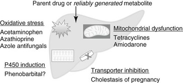
Drug-Associated Liver Disease

Chapter 140


Drug-Associated Liver Disease*


The liver is a common target of drug toxicity. It receives 25% to 30% of cardiac output and is the site of first-pass clearance of many orally administered drugs. Hepatic cytochrome P450s and other biotransformation enzymes can generate reactive metabolites, which may lead to local cytotoxicity, or haptenize liver proteins and trigger an immune response. Drugs also can inhibit transporter pumps in the sinusoidal or canalicular membranes, thus interfering with hepatocyte function and bile salt efflux. Two major patterns of drug-induced hepatotoxicity are recognized: cytotoxic (associated with hepatocyte necrosis) and cholestatic (which can be attributed to inhibition of biliary transporters, or mitochondrial injury with resulting steatosis). In humans, an R value is calculated to characterize hepatotoxicity by biochemical pattern, where


image


An R value greater than 5 indicates hepatocellular injury, an R less than 2 indicates cholestatic injury, and an R of 2 to 5 represents a mixed pattern.


Dose-Dependent Hepatotoxic Drugs


For dose-dependent, or intrinsic, hepatotoxicity, toxicity increases with increasing dose in one or more species, and virtually all members of a population or species are affected at high enough doses. Dose-dependent hepatotoxicity may be caused by the parent drug or by a metabolite generated reliably in the treated species (Figure 140-1). These reactions are relatively predictable, and therapeutic drug monitoring may be helpful in prevention. They require a dose reduction but usually not permanent drug discontinuation.


Phenobarbital


Hepatotoxicity from phenobarbital can range from subclinical increases in serum bile acids to clinical hepatopathy to fulminant liver failure (Dayrell-Hart et al, 1991). Signs typically develop after a year or more of phenobarbital treatment, and prolonged duration of administration is associated with degree of histologic injury in epileptic dogs. Typical histologic findings in dogs with clinical signs are bridging portal fibrosis, bile duct hyperplasia, and nodular regeneration. Dogs with phenobarbital hepatotoxicity improve clinically after phenobarbital dose reduction (Dayrell-Hart et al, 1991). However, liver enzyme abnormalities can develop in phenobarbital-treated dogs without histologic liver injury, and higher phenobarbital dosages and serum drug concentrations have not been correlated with the development of abnormal serum bile acids in epileptic dogs. These findings suggest that phenobarbital hepatotoxicity is dose dependent, with modifying factors that are not understood.


One hypothesized mechanism of phenobarbital hepatotoxicity is induction of cytochrome P450 enzymes, with secondary bioactivation and hepatotoxicity of other drugs, dietary components, or environmental toxins. For example, phenobarbital increases the hepatotoxicity of carbon tetrachloride in dogs, of chloroform in mice, and of acetaminophen in human hepatocytes. Phenobarbital hepatotoxicity therefore could be modulated by environmental exposures in individual dogs. Phenobarbital does not lead to either enzyme induction or hepatotoxicity in cats.


Azathioprine


Azathioprine can lead to increases in ALT and/or ALP activities in some dogs; these abnormalities commonly are subclinical but may be accompanied by jaundice and clinical signs. Azathioprine liver injury is associated with the generation of oxidative metabolites and depletion of hepatic antioxidants in rodent models and can be prevented by pretreatment with N-acetylcysteine.


Dogs treated with azathioprine should be monitored routinely for increases in liver enzyme activities. If glucocorticoids also are administered, liver enzyme interpretation can become clouded; discordant increases in ALT relative to SAP, or early increases in serum bilirubin, are cause for concern. Risk factors for azathioprine hepatotoxicity in dogs are not clear, but increases in ALT or ALP typically are reversible with simple dosage reduction. Based on what is known about azathioprine hepatotoxicity in other species, supplementation with glutathione precursors may be effective in preventing or reversing azathioprine hepatotoxicity in dogs, but this has yet to be evaluated.


Azole Antifungals


Ketoconazole, itraconazole, and fluconazole can lead to increases in serum ALT in dogs, although clinical signs such as jaundice are uncommon. Mild, clinically insignificant increases in ALT also have been reported in cats treated with itraconazole and fluconazole. In dogs with blastomycosis, higher dosages of itraconazole (10 mg/kg/day) were associated with a greater risk of ALT abnormalities than 5 mg/kg/day, with no difference in efficacy (Legendre et al, 1996). Further, increases in ALT and ALP were correlated with itraconazole plasma concentrations, which supports a dose-dependent mechanism.


In animal models, ketoconazole hepatotoxicity has been attributed to an oxidative metabolite, N-deacetyl ketoconazole, which leads to covalent binding to liver proteins and glutathione depletion. Fluconazole appears to be less hepatotoxic overall than either ketoconazole or itraconazole in humans and animal models. In dogs with blastomycosis, the author observed increases in serum ALT in 26% of dogs treated with itraconazole (median fold increase 2.7), and in 17% of dogs on fluconazole (median fold increase 1.5). The author has observed anecdotally increases in serum ALT during treatment with itraconazole, which have resolved after a switch to fluconazole during treatment for blastomycosis in dogs.


Acetaminophen


Acetaminophen is a classic dose-dependent hepatotoxin in humans and dogs; doses greater than 150 to 250 mg/kg lead to acute centrilobular hepatic necrosis. In cats, hematologic toxicity predominates over direct liver toxicity. Acetaminophen is bioactivated to the reactive oxidized metabolite, NAPQI (N-acetyl-p-benzoquinone imine), which is detoxified by glutathione conjugation. This provides the rationale for treatment of overdoses with the glutathione precursor N-acetylcysteine (140 mg/kg loading IV, then 70 mg/kg q6h for 7 treatments). Although N-acetylcysteine is most effective in humans when given within 8 hours of acetaminophen ingestion, this antidote still has beneficial effects on survival when given much later in the course of intoxication.


S-adenosylmethionine (SAMe) also can be used for acetaminophen intoxication in dogs that can tolerate oral medications; the protocol that has been used successfully is a 40 mg/kg loading dose, followed by 20 mg/kg q24h for 7 days. Cimetidine has been recommended to inhibit oxidation of acetaminophen to NAPQI; however, this drug has no effect on NAPQI generation in vitro and is not effective in humans with acetaminophen overdose; cimetidine therefore is not recommended.


Amiodarone


Amiodarone leads to clinically significant hepatotoxicity in about 45% of dogs treated for refractory atrial fibrillation and ventricular arrhythmias, a median of 16 weeks after starting maintenance therapy (Jacobs et al, 2000; Kraus et al, 2009). Predominant increases in ALT typically are observed, with or without hyperbilirubinemia and neutropenia. These abnormalities slowly resolve over 1 to 3 months after drug discontinuation. Toxicity in animal models has been attributed to two oxidative metabolites, mono-N-desethylamiodarone (MDEA) and di-N-desethylamiodarone (DDEA), which generate reactive oxygen species that uncouple oxidative phosphorylation and lead to mitochondrial damage.


Because of the prevalence of hepatotoxicity and neutropenia, a baseline CBC and biochemical panel is recommended in all dogs before amiodarone initiation, with a recheck of liver enzymes after a loading period and monthly during treatment. The development of substantial increases in serum ALT is an indication for dose reduction or drug discontinuation.

< div class='tao-gold-member'>

Stay updated, free articles. Join our Telegram channel

Jul 18, 2016 | Posted by in PHARMACOLOGY, TOXICOLOGY & THERAPEUTICS | Comments Off on Drug-Associated Liver Disease

Full access? Get Clinical Tree

Get Clinical Tree app for offline access