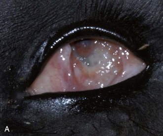
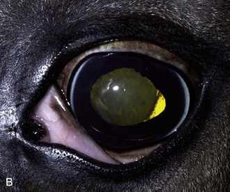
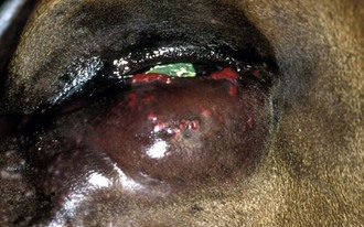
Chapter 3 Diseases and Surgery of the Globe and Orbit
Clinical Anatomy and Physiology
Skull and Foramina
The horse is unusual in having a complete bony orbital rim (Fig. 3-1). The orbit and globes are positioned laterally on the head and are ideally situated for a wide and panoramic visual field exceeding 340 degrees.1 This seems appropriate for the horse’s ecologic niche as a grazer and consequently a prey animal, but perhaps it is less perfect for the activities it is commonly tasked in today’s society. The visual field is binocular, permitting stereoscopy, and the wide separation of the two globes provides greater depth perception than most other domestic species can attain. The main visual axis is approximately parallel with the long axis of the skull.2 Equine vision is explored more thoroughly in Chapter 11.
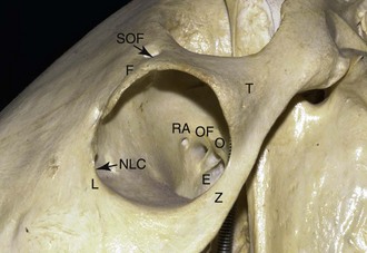
Figure 3-1 The horse has a complete bony orbital rim. The orbital bones include the frontal (F), lacrimal (L), zygomatic (Z), and temporal (T). The sphenoid and palatine bone forming the medial wall of the orbit separates the orbit from the calvarium. The nasolacrimal duct canaliculi (NLC) course through the lacrimal bone. The orbital foramina through the sphenoid bone includes, from anterior to posterior, the rostral alar (RA), orbital fissure (OF), optic (O), and ethmoidal (E) (see Table 3-1). The supraorbital foramen (SOF) exits through the frontal bone.
The equine globe is among the largest of the mammalian kingdom, perhaps eclipsed in size only by the giraffe’s eye. The axis of the globe in the horse is nearly perpendicular to that of the skull and angled with respect to the long axis of the orbit. The average dimensions of the adult horse globe are 43.68 mm anterior-posterior, 47.63 mm vertically, and 48.45 mm horizontally.3 The globe of the mule is almost identical in size, measuring 43.0 mm, 47.5 mm, and 48.5 mm in the same orientation. The globe is not a sphere, but rather is compressed from anterior to posterior, with the ratios of the dimensions previously given being 1:1.09:1.10 for the horse and 1:1.10:1.12 for the mule.3 The globe is located at the anterior border of the rather large orbit, which averages 62 × 59 × 98 mm. The orbits are separated by 172 mm across the anterior surface of the frontal bone.3 The remainder of the orbital cavity is filled largely by fat and muscle and is divided into pockets by layers of connective tissue that coordinate ocular mobility.
The orbital bones are the frontal, lacrimal, zygomatic, and temporal bones (clockwise for the right orbit), with the sphenoid and palatine bone forming the deeper internal wall of the orbit that separates it from the calvarium (see Fig. 3-1). The complete bony rim temporally and dorsally is composed of the frontal process of the zygomatic bone and zygomatic processes of the frontal and temporal bones. The supraorbital foramen is located in the anterior aspect of the frontal bone that forms the dorsal orbital rim. The lacrimal gland is closely adhered to the internal orbit near the dorsotemporal orbital rim. Inferiorly the zygomatic bone forms the lower rim, and nasally it is fused to the lacrimal bone, wherein embedded canals provide refuge to the nasolacrimal duct canaliculi (see Fig. 3-1). Beneath the globe, cushioning is provided by orbital fat that separates the globe from the palatine bone. Caudally the orbital floor is angled inferiorly until it is composed of soft tissues only, including the large pterygoid muscles that exert closing motion for the lower jaw. The bony floor of the orbit ends directly beneath the caudal aspect of the dorsal rim.
Various foramina provide conduits between the orbit and other compartments of the head, particularly through the calvarium to the brain. The efferent foramina are arranged curvilinearly along the sphenoid bone that forms the medial orbital wall. From anterior to posterior they are the rostral alar, orbital fissure, optic, and ethmoidal (Table 3-1) and are evident in Fig. 3-1. The clinical significance of the location of the foramina lies in avoiding their contents during biopsy and exploratory procedures. The foramina appear to provide relatively minimal risk for extension of orbital infection or neoplasia, but they may be severely damaged during trauma to the calvarium, with serious consequences for vision. Fracture of the basisphenoid and basioccipital bones frequently occurs in blunt trauma to the poll. Afferent foramina are located in the extreme anteromedial portion of the orbit; they are the caudal palatine (major palatine artery and nerve), the maxillary (infraorbital artery, vein, and nerve), and sphenopalatine (sphenopalatine artery and vein and pterygopalatine nerve).
Table 3-1 | Structures Perforating the Orbit and Contents
| STRUCTURE | OSTEUM |
|---|---|
| Maxillary artery and nerve | Rostral alar foramen |
| Ethmoidal artery, vein, nerve | Ethmoidal foramen |
| Optic nerve (II) | Optic foramen |
| Oculomotor nerve (III) | Orbital foramen |
| Trochlear nerve (IV) | Orbital foramen |
| Abducens nerve (VI) | Orbital foramen |
| Supraorbital artery, vein, nerve | Supraorbital foramen |
| Major palatine artery, vein, and nerve | Caudal palatine foramen |
| Infraorbital vein, artery, and nerve | Maxillary foramen |
| Sphenopalatine artery and vein and pterygopalatine nerve | Sphenopalatine foramen |
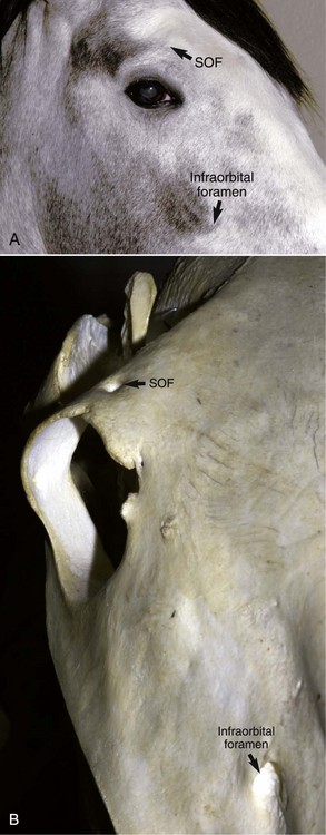
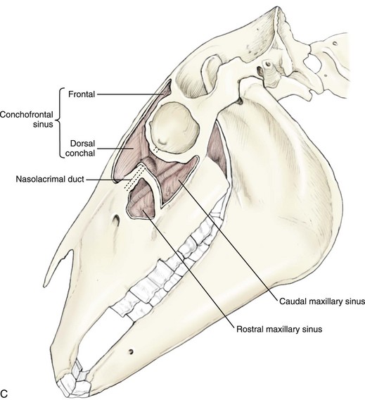
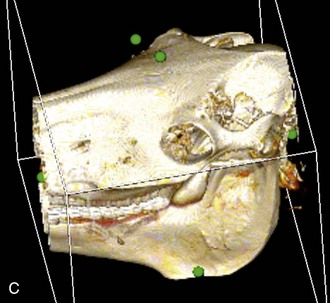
The infraorbital foramen is the point of egress of the infraorbital nerve, artery, and vein from the orbit, which can be palpated 1 cm rostral and 3 cm dorsal to the rostral edge of the facial crest, a third of the distance to the nostrils (Fig. 3-2, A and B). It may be partially obscured by the overlying muscular belly of the levator nasolabialis.4 The infraorbital nerve arises from the maxillary branch of the trigeminal to provide sensory innervation to the upper lip, nostril, and cheek. Its major significance is in desensitization for laceration repair of those tissues, and infrequently for nerve blockade to evaluate the contribution of trigeminal summation in clinical head shakers.5 The infraorbital canal bisects the maxillary sinus (see Fig. 3-2, C).
Globe
The equine globe resides anteriorly within the orbit, supported by loosely packed retrobulbar tissues. Reflections of the periorbitum together with smooth muscle attach the globe to the orbital rim. The external wall of the globe is a trilayer, with the sclera and cornea forming the outer predominantly fibrous tunic, the highly vascular uvea forming the middle layer, and the neural layers of the retina being most internal. The thickness of the sclera is 1.05 mm where the optic nerve penetrates the posterior pole, 0.31 mm at the limbus, and 0.5 mm at the equator of the globe.6 Consequently, during traumatic events, the sclera most frequently ruptures at the limbus, the potentially weakest area of the sclera. The adult cornea measures 29.7 to 34.0 mm horizontally and 23.0 to 26.5 mm vertically.7 When measured by ultrasonic pachymetry, the corneal thickness averages 0.793 to 0.893 mm, with no effect of gender or age but a tendency to be thicker in the dorsal and ventral quadrants.8,9 Recently these values have also been reported for Miniature horses, where average corneal thickness was 0.785 mm, horizontal diameter was 25.8 mm, and vertical diameter was 19.4 mm.10 Mean intraocular pressure was 26.0 mm Hg.10 The internal volume of the equine globe is large. In one study, the mean aqueous humor volume measured in six adult equine eyes was 3.04 ± 1.27 mL, and the mean vitreous humor volume was 26.15 ± 4.87 mL.6 Total internal volume of the globe has been reported up to 45 to 50 mL with a weight of 100 mg.6
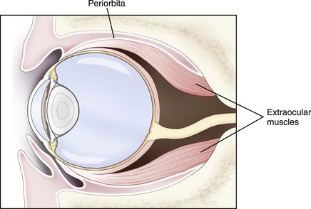
The multiple large bellies of the retractor bulbi muscle, which attach extensively around the posterior half of the sclera and are innervated by cranial nerve VI, dominate the extraocular muscles (Fig. 3-3). The globe retraction reflex provides one of the major methods of evaluating this nerve’s function. The other six muscles have typical attachments (Table 3-2) but more extensive bulk and insertion than is the case in many other species. Muscular attachments are seldom avulsed in orbital trauma (in contrast to the dog), but they may become entrapped and be unable to function after orbital fractures.
Table 3-2 | Orbital and Ocular Muscles: Function and Innervation
| EXTRAOCULAR MUSCLE | FUNCTION | INNERVATION |
|---|---|---|
| Dorsal rectus | Upward motion of globe | Oculomotor (III) |
| Ventral rectus | Downward motion | Oculomotor (III) |
| Medial rectus | Medial motion | Oculomotor (III) |
| Lateral rectus | Lateral motion | Abducens (VI) |
| Dorsal oblique | Rotation nasally and inferiorly | Trochlear (IV) |
| Ventral oblique | Rotation laterally and superiorly | Oculomotor (III) |
| Retractor bulbi | Posterior motion of globe | Abducens (VI) |
| EYELID | FUNCTION | INNERVATION |
|---|---|---|
| Levator palpebrae superioris | Elevates upper eyelid | Facial (VIII) |
| Levator angularis oculi | Elevates nasal eyelid/eyebrow | Oculomotor (III) |
| Malaris | Opens lower eyelid | Facial (VII) |
| Orbicularis oculi | Forceful closure of eyelids | Facial (VII) |
| Retractor angularis oculi | Retracts lateral canthus | Facial (VII) |
| Arrectores ciliorum | Elevates eyelashes | Sympathetic |
| Orbitalis (Muller’s): superior and inferior tarsus and circular fibers | Retracts eyelid (smooth) | Sympathetic |
| Corrugator supercilii | Elevates upper eyelid | Facial (VII) |
Orbital Vascular Supply
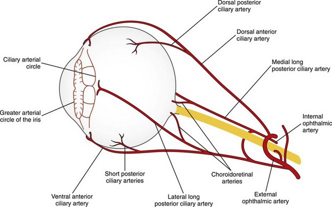
The vascular system of the equine orbit has been described in detail, and the reader is referred to the excellent work of Simoens.11 Most vessels cross through the orbit inferiorly and nasally and may be avoided by aspirating and biopsying the orbit only from the dorsal, lateral, or dorsomedial directions. The orbital branches of the external ophthalmic artery are variably derived from the internal maxillary artery deep within the orbit (Fig. 3-4). Two functional groups—the ciliary arteries rostrally and the chorioretinal arteries caudally—perfuse the globe. Posterior ciliary arteries enter the sclera behind the equator in the cardinal positions to perfuse the choroid and continue forwards toward the anterior segment within the sclera. Anterior ciliary arteries enter superiorly and inferiorly caudal to the limbus. Multiple chorioretinal arteries (10 to 20) surround the optic nerve at its entry through the sclera, supplying retinal arteries and contributing to local choroidal perfusion.11 The primary venous drainage of the eye is through the ophthalmic, orbital, supraorbital, and reflex veins.3 The superior vortex vein, palpebral, and lacrimal veins course into the dorsal ophthalmic vein before more caudal contributions from the anterior ciliary, supraorbital, muscularis, and infratrochlear veins, and ultimately from the ophthalmic vein at the apex of the orbit. The orbital and internal maxillary vein then provides venous drainage. The inferior vortex vein anteriorly drains into the reflex vein, which also receives venous return from the muscular and palpebral veins, the great palatine, sphenopalatine, and infraorbital veins in the anterior orbit before entering the facial vein.
Structures Adjacent to the Orbit
Guttural Pouch
The guttural pouch is a diverticulum of the eustachian tube that occupies the area posterior to the mandible and is the site of multiple fragile structures. It extends anteriorly to closely approximate the orbit and enters the pharynx through a slitlike opening. Therefore, disease of the guttural pouch may directly impact the orbit. More commonly, ocular disease may result from damage to the internal carotid artery that passes through the guttural pouch or to the cranial cervical ganglion of the autonomic nervous system. The major diseases of the guttural pouch are bacterial or fungal infection and empyema, and some of the surgical and medical managements of that condition. Rupture of the internal carotid artery is a life-threatening condition. See Chapter 13, Ocular Manifestations of Systemic Disease, for more information on guttural pouch disease.
Nasolacrimal Duct
The nasolacrimal duct system originates proximally via large puncta in the upper and lower eyelids and is joined by canaliculi to the lacrimal sac, which is embedded in the lacrimal bone. Damage by fracture, direct blunt trauma, infectious or inflammatory disease at this site may cause epiphora and chronic irritation. Treatment of nasolacrimal disease is described in Chapter 4. The nasolacrimal duct traverses the maxillary sinus, and sinusitis resulting in increased sinus pressure may functionally obstruct the duct, resulting in epiphora.
Periorbital Sinuses
Periorbital sinuses, the frontal (conchofrontal), maxillary (caudal and rostral), and sphenopalatine, are in close anatomic proximity to the orbit (see Fig. 3-2, C). Primary sinus diseases may secondarily affect one or both orbits.12–16 The frontal sinus is a shallow, wide sinus parallel with the frontal bone; it extends anteriorly from a line joining each temporomandibular joint forward almost to the dorsal turbinate bone that represents the conchofrontal sinus. The sinus system is lined with epithelium and is air filled, therefore an open fracture of any sinus is clinically relevant because it is considered a contaminated wound. The maxillary sinus extends ventrally from an imaginary line joining the medial canthus and the nasomaxillary notch, to just below the facial crest. The rostral border is the rostral extent of the facial crest, and the caudal border is the midline of the orbit. Diminutive ethmoidal and sphenopalatine sinuses are present medial to the internal wall of the orbit and are more difficult to identify from external landmarks. Infection of the sphenopalatine sinuses may cause swelling and distention that can compress the optic nerve and chiasm, resulting in blindness.17 Together with the frontal sinus, drainage is into the caudal maxillary sinus. Ultimately, drainage occurs from these sinuses into the nasal cavity. When surgical drainage is indicated for sinus disease, trephination dorsal to a line between the infraorbital foramen and the medial canthus can result in nasolacrimal duct damage and must be avoided (see Fig. 3-2, C).18
Orbital Diagnostics
Orbital Imaging
Orbital disease may be more challenging to diagnose because many of the structures are hidden from direct examination. In addition to appearance, displacement of adjacent structures permits deduction of involved structures and suggests the tissue of interest. Imaging systems provide confirmation of suspected diagnoses and are frequently an essential part of the diagnostic process. Imaging is described in greater detail in Chapter 1.
Ultrasonography
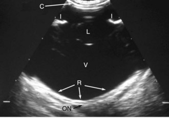
Ultrasonography is convenient, efficient, performed in real time, inexpensive, and readily available to most practitioners.19,20 Although a 10- or 7.5-MHz probe is necessary for adequate imaging of the globe, the deeper aspects of the orbit may require a 5- or 7.5-MHz probe (Fig. 3-5). However, orbital ultrasonography is not very sensitive or specific, and other imaging modes such as CT may be more diagnostic. In fact, even moderately large orbital lesions may not be apparent on some ultrasound examinations. Please see Chapter 1 for the technique on how to perform the ocular and orbital ultrasound.
Aspiration of a lesion (e.g., mass, fluid) in the equine orbit can be performed for cytology, culture, and histopathologic examination. An 18-gauge, 10-cm, slightly curved needle is inserted 1 cm lateral to the lateral canthus and then directed posteriorly in a line parallel to the medial canthus.21 Approaching the retrobulbar space via the supraorbital fossa as described for retrobulbar block is also possible, especially if the lesion is in the dorsal retrobulbar area.
Exophthalmos and orbital trauma are the two most common indications for orbital ultrasound examination in the horse. After trauma to the orbit and associated structures, ultrasound may be used to evaluate the retrobulbar space for displaced fractures, hemorrhage, swelling, compression of the optic nerve, and integrity of the posterior wall of the globe.22
Radiography
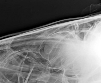
Radiography is indicated primarily to identify bone fractures in the acutely traumatized individual, bone deformation in invasive neoplasia or sepsis, and to localize cellulitis if gas production is evident. Precise radiographs require general anesthesia to avoid motion, although screening images may be obtained in the standing sedated patient, particularly with the advent of digital radiography. Specific skyline techniques to highlight the orbital bones of interest (Fig. 3-6) may identify a lesion against an air-filled background (e.g., frontal sinus). Contrast orbitography may be performed by artificially introducing air into the orbit, although routine CT and MRI are much more sensitive diagnostic procedures. Other injectable contrast materials are seldom indicated and risk creating complications. Fluid/air interfaces within the sinus indicate pathology that is directly relevant to the orbit.
Computed Tomography and Magnetic Resonance Imaging
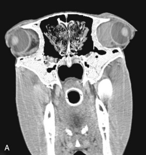
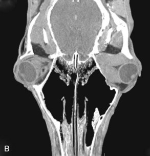
Advanced imaging such as MRI and CT provide the best images to localize the orbital lesion precisely (Fig. 3-7). The use of cross-sectional and three-dimensional reconstruction of CT equine skull images overcomes the superimposition of the complex anatomic features of the equine skull that make conventional radiographic interpretation difficult.23,24 The large amount of low-density fat in the equine orbit provides good natural contrast for differentiating various soft-tissue structures by CT, although the primary advantage of CT over MRI is its ability to detect osseous changes.23 Disadvantages of CT over skull radiography in the horse include the need for general anesthesia and the risks associated with recovery from anesthesia, increased cost, and limited accessibility.24 There have been several reports of CT examination of the equine orbit, including evaluation of the periorbital sinuses, orbital masses, and skull fractures.17,24–26 See Chapter 1 for more information on CT imaging.
Impact of Orbital Diseases on the Equine Industry
There are no significant contagious or infectious diseases of the orbit that pose a threat to livestock in the United States. Among exotic diseases, African horse sickness, equine infectious anemia, and infectious diseases causing vasculitis may cause retrobulbar and supraorbital swelling and distension in multiple individuals (see Chapter 13). Quarantine, importation restrictions, and routine serologic testing are the major source of protection against these diseases in the United States, Australia, and nonendemic areas of Europe.
Clinical Signs of Orbital Disease
Abnormalities of Globe Position
Strabismus is the deviation of the visual axis from the expected orientation, which is straight ahead in the horse. When both globes are focused ahead, the conjugate gaze has an overlap of the visual fields, allowing for binocular vision and good depth perception owing to the wide globe separation (see Chapter 11). The deviation induced by strabismus presumably disturbs the collation and overlap of visual field information from each eye when it is interpreted in the midbrain and visual centers. In people, misdirected visual axes result in diplopia or double vision. Strabismus generally occurs from a space-occupying mass of the orbit, distorting the globe from its resting position, or possibly it may result from disturbance of the ocular muscle innervation or activity due to stricture, contraction, or mechanical entrapment. Strabismus in young individuals is uncommon and likely to be congenital or breed related.
Neonatal foals most frequently have a physiologic rotational strabismus that resolves to the adult orientation within the first 2 to 4 weeks of life. The horizontal pupil is rotated so that the nasal portion is ventral to the temporal portion, and the globe may also be rotated medially. The strabismus is considered physiologic and requires no therapy. The cause is unknown, and no therapy is possible. Appaloosa foals afflicted with congenital stationary night blindness (CSNB) may also exhibit a dorsal strabismus (Fig. 3-8).27,28 The relationship of the strabismus to CSNB is unclear.
Acquired causes of strabismus are traumatic in origin typically, due to muscle entrapment post fracture, or less likely due to avulsion of a rectus muscle attachment. The most anterior rectus muscle attachments are for the ventral (8 mm behind limbus) and dorsal (9 mm) rectus muscles, while a portion of the lateral rectus attaches within 5 mm of the limbus.29 Occasionally, central nervous system infections may result in strabismus, most commonly with equine protozoal myeloencephalitis (see Chapter 13).30–32 Midbrain (oculomotor) lesions most commonly result in a lateral strabismus.33 Peripheral vestibular disease may result in strabismus rather than nystagmus and is often accentuated by elevation of the horse’s head. Strabismus may be the only residual sign after recovery from vestibular disease.33
Nystagmus
Nystagmus is an oscillatory movement of the globe which may be horizontal, vertical, or rotational. Physiologic nystagmus is a normal compensatory reaction that occurs when the head turns and is a direct response to differential stimuli within the inner ear and the vestibular system. This involuntary motion of the globe permits a more stable visual horizon and enhances stability of the visual field. Pathologic forms of nystagmus reflect damage to the vestibulocochlear apparatus (horizontal, rotatory) or central nervous system disease (vertical). Nystagmus most frequently accompanies central disease in the horse, which is most often infectious in etiology. Potential etiologies include equine protozoal myeloencephalitis (EPM), the viral encephalitides (Eastern equine encephalitis [EEE], Western equine encephalitis [WEE], Venezuelan equine encephalitis [VEE], West Nile Virus), aberrant parasite migration, or bacterial sepsis. Peripheral causes include EPM; trauma fracturing the petrous temporal, basisphenoid, or basioccipital bones; conditions afflicting the guttural pouch diverticulum (empyema, mycosis, chondroids); temporohyoid osteopathy (THO); and osteomyelitis adjacent the inner ear and orbit. Concurrent depression substantially worsens the prognosis. Refer to Chapter 13 for more details of the specific diseases and their treatment.
Abnormalities of Globe Location, Size, and Function
Exophthalmos
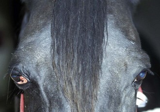
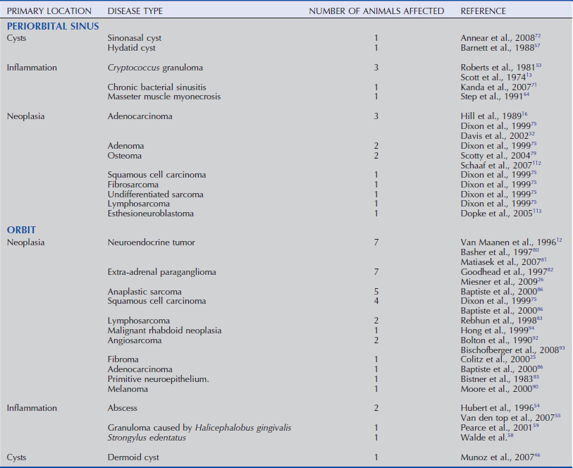
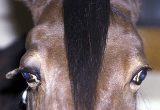
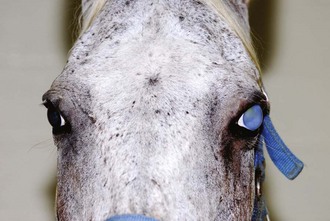
Exophthalmos describes the anterior displacement of a normal-sized globe within the orbit. The most common causes are retrobulbar masses, orbital cellulitis, and trauma that reduces the orbital space (Fig. 3-9 and Table 3-3). Exophthalmos is most easily identified by viewing the head from the front and comparing the angle of eyelashes, the relative prominence of the globe, and the size of the palpebral fissure (Fig. 3-10). In contrast to other domestic species, there is minimal additional benefit from observation from above the skull. An exophthalmometer is a device used to measure relative globe prominence within the orbit, but it is seldom of clinical relevance. Palpation of the globe relative to the orbital rim may achieve similar results to an exophthalmometer.
Enophthalmos
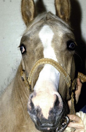
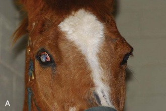
Enophthalmos, or globe abnormally deep or caudally displaced in the orbit of the globe, is typically secondary to the loss of orbital contents. It is most commonly found in older horses that are reabsorbing orbital fat, but it should be differentiated from phthisis bulbi occurring in a chronically inflamed eye. Neonatal foals are enophthalmic if dehydrated (Fig. 3-11) and may develop entropion due to lack of globe support to the eyelids. This neonatal foal enophthalmos reverses rapidly with return to normal homeostasis. Enophthalmic globes should be comfortable and visual, but vision may be reduced by the prominence or elevation of the nictitans, which is no longer held in position by the globe. The supraorbital fossa is more prominent and deep. It has been suggested that fat mobilization occurs preferentially from sites with the greatest variation in individual fat-cell size. The orbital fat and supraorbital fossa fat cells are the smallest in the body, suggesting that fat resorption in this area occurs as a late outcome.34
Trichiasis and chronic ocular discharge from misalignment of the eyelid and puncta are management problems in enophthalmos. Entropion of the lower eyelid may be addressed to prevent direct abrasion of the hair on the cornea. (See Chapter 4 for more information on management of entropion and trichiasis.) Strategies include primary entropion repair, which may result in inappropriate gapping between the globe and the eyelid and accumulation of mucoid debris. Alternatives are cryosurgery of irritating hairs, injection of collagen intradermally to increase eyelid rigidity, or placement of retrobulbar silicone devices to propel the globe forward. The latter procedure is subject to later displacement of the silicone prostheses and recurrence. Addressing the sequelae in this manner is likely to only be temporarily successful. Pars intermedia dysfunction in horses may accentuate reabsorption of fat and worsen enophthalmos. Orbital fracture may result in enophthalmos if the ventral floor of the orbit is displaced. Finally, enophthalmos may be associated with sympathetic denervation to the smooth muscle between the globe and orbital rim, which is typically somewhat subtle in appearance but may be associated with nictitans prominence (see Chapters 4 and 13 for more information on Horner’s syndrome).
Phthisis Bulbi
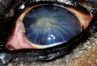
Phthisis bulbi, gradual shrinkage of the globe, is due to chronic inflammation and hypotony of the globe resulting from severe damage to the ciliary body epithelial cells responsible for fluid production (Fig. 3-12). Generally, eyes that are phthisical should be enucleated to prevent ocular discomfort. When a cosmetic appearance is desirable, phthisical globes may have an intrascleral prosthesis inserted to replace the intraocular contents. It is not possible to expand the size of the globe, but further shrinkage may be avoided. A cosmetic shell with or without enucleation and orbital prosthesis placement may also result in reasonable cosmetic outcomes (see technique later in chapter).35 See chapters on uveal diseases (see Chapter 6) and equine recurrent uveitis (see Chapter 8) for more information and treatment of intraocular inflammatory diseases.
Atrophia Bulbi
Atrophia bulbi describes a smaller globe than normal, suggesting a gradual decline in globe size secondary to chronic inflammation. In contrast, phthisis bulbi refers to an end-stage, blind eye. Again, Chapters 6 and 8 offer more information on diagnosis and treatment of intraocular inflammatory diseases.
Hydrophthalmos (Buphthalmos)
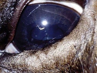
Hydrophthalmos refers to an unusually enlarged globe, which is associated with chronically increased intraocular pressure secondary to glaucoma. Technically, the term buphthalmos indicates a globe typical of a bovine, which purists would observe is generally smaller than that of the horse. Hydrophthalmos indicates that the globe is larger than normal and may be the preferred term in the horse. In adult horses, globe enlargement occurs slowly and is associated with other clinical signs of glaucoma, including corneal edema and endothelial striae (Fig. 3-13). Vision may be present, reduced, or absent at the time of diagnosis. It is not uncommon in horses for vision to return when intraocular pressure (IOP) is subsequently controlled, even if the condition is chronic and mild globe enlargement has occurred. Glaucoma may also be congenital, in which instance hydrophthalmos may occur rapidly with only a temporary retention of ocular function. Congenital glaucoma is usually severe and blinding in the early stages. Hydrophthalmos should be differentiated from exophthalmos of a normal-sized globe. See Chapter 9 for more information on the diagnosis and treatment of glaucoma in horses.
Megalophthalmos
Megalophthalmos refers to a distortion of the globe and has been used to describe the grouping of abnormalities found in Rocky Mountain horses with multiple congential ocular anomalies. These clinical signs include increased corneal curvature, iris hypoplasia, congenital miosis, uveal cysts, cataracts, and retinal dysplasia. Persistent pupillary membranes may also be present.36
Other Signs of Orbital Disease in the Horse
Prominent Nictitans
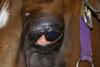
The nictitans in the horse can be elevated passively with orbital space-occupying masses (Fig. 3-14). This temporary physiologic displacement may become chronic and persistent if orbital contents are altered in position due to a mass or absence of adequate globe support resulting in enophthalmos. The prominence of the nictitans in Horner’s syndrome is associated with enophthalmos and may not always be obvious in the horse. Clinical cases of tetanus often exhibit spasm of the nictitans, particularly in response to sudden stimulation. Globe retropulsion should always be performed when nictitans prominence is observed.
Epiphora
Epiphora is commonly associated with orbital disease, but it should be differentiated from eyelid swelling and distortion such as that due to dacryocystitis, neoplasia, or infection (see Chapter 4). In orbital and extraorbital disease, the nasolacrimal duct may be obstructed functionally or anatomically (i.e., by sinusitis, fracture, or neoplasia), or the eyelid puncta may become misaligned due to blepharoedema, blepharoconjunctivitis, or exophthalmos. Normal tear flow through the nasolacrimal duct depends in part upon vacuum development in the duct, created by the orbicularis oculi during eyelid closure. Functional obstruction may be differentiated from occlusion by irritation of the nasolacrimal duct.
Emphysema
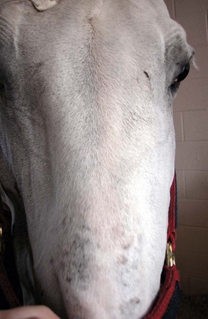
Accumulation of subcutaneous air is due to unidirectional aspiration of air and entrapment between the skin and calvarium (Fig. 3-15). The most common cause is a sinus fracture, which should be considered an open contaminated wound. A depression or displaced fracture is almost always palpable, but radiography with skyline views or a CT scan should be performed if the fracture is not identified clinically. Infrequently, anaerobic bacterial sepsis may result in gas production, although this is uncommon in the equine head.
Episcleral Vessel Enlargement
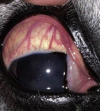
If direct compression of the retrobulbar optic cone results in venous stasis, episcleral vessels may become quite prominent (Fig. 3-16). Elevated intraocular pressure, or glaucoma, may also develop and it is typically poorly or unresponsive to hypotensive medications, despite the importance of uveoscleral outflow in the horse. Exophthalmos may or may not be present. Retropulsion may not be abnormal because the functional obstruction surrounds the cone in the caudal orbit. Blindness may result from concurrent optic nerve compression, even if retropulsion is still possible.
Keratoconjunctivitis Sicca
Keratoconjunctivitis sicca together with ipsilateral facial nerve paralysis suggest trauma to the facial nerve proximal to the geniculate ganglion. Neuroparalytic keratitis results from more distal lesions. The vestibulocochlear nerve (VIII) should be evaluated critically if facial (VII) nerve disease is identified, because they are commonly injured together in proximity to the petrous temporal bone29 or the ramus of the mandible.37
Supraorbital Fossa Distension
Supraorbital fossa distension may accompany exophthalmos and space-occupying masses of the orbit (see Fig. 3-10). It is a particularly prominent feature of the exotic disease African Horse Sickness, where hemorrhagic edema causes great distension. Equine viral arteritis results in a panvasculitis and secondary edema that appears similarly (see Chapter 13 for further details).
Congenital Diseases
Microphthalmos
Microphthalmos is a congenital or developmental anomaly in which the globe is abnormally small.20,38–43 Microphthalmos may be simple or complicated, referring to whether it has compound abnormalities or not. Embryologically, microphthalmos may result from incomplete closure of the optic fissure, preventing normal sealing and inflation of the globe, or it may be the consequence of a mistimed union of the migrating embryologic tissues. Microphthalmia occurred in 14.7% of foals in one study39 and 7% in another.44 A case of microphthalmia with brachygnathia has been reported in an older Friesian mare that received griseofulvin in the second month of gestation, similar to microphthalmia due to intoxication in other breeds.42 Multiple ocular colobomas may accompany microphthalmia and result in severely debilitated and blind globes.
Clinical Appearance and Diagnosis
Microphthalmic eyes appear variably smaller than normal (Fig. 3-17) and commonly have additional abnormalities including cataract and other congenital abnormalities such as retinal coloboma. Nanophthalmos refers to a globe that is functional but smaller than normal. Anophthalmos is the furthest degree of microphthalmos and is extremely uncommon. More commonly, it is an excessively small globe that is barely recognizable and usually present as a very diminutive and heavily pigmented spherical mass. In such cases, the predominant tissues identified are conjunctivae and nictitans. In the adult animal, differentiating mild microphthalmos from phthisis bulbi as the cause of the small globe may be difficult; however, a poorly developed, small orbit and historical reports of a small or absent globe are most common with microphthalmos. Phthisical globes typically exhibit intraocular changes suggestive of prior substantial uveitis (see Chapters 6 and 8).
Treatment
If the lens is cataractous but the globe approaches normal size, has appropriate light responses, and a normal electroretinogram, it is often possible to restore useful vision with the removal of the cataract if surgery is performed early (see Chapter 7). If practical management is a substantial issue and the condition is unilateral, the globe may be removed and possibly replaced with a prosthetic device (see later). For a more normal development of the orbit, stepwise increase in orbital conformer size may assist in the approximation of normal orbit size in the adult horse.
Congenital Glaucoma
Congenital glaucoma occurs uncommonly in foals but is often quite dramatic. The globe may rapidly enlarge and become a management problem before it becomes blind. Therapy usually carries a poor prognosis for maintaining vision. (For more information see Chapter 9.)
Orbital Dermoids
Orbital dermoids are particularly rare in horses but may cause congenital or juvenile unilateral exophthalmos. Skin with or without hair embedded within the orbit may result in a fluid-filled cystic mass that expands.41,45,46 In one report, exophthalmos did not develop until the horse was 4 years old.46 More commonly, noncystic dermoids are located in the dorsotemporal conjunctiva or limbus; they are frequently pigmented but not always haired.
Clinical Appearance and Diagnosis
Clinical presentation is as for any other space-occupying mass in the orbit. Progressive enlargement and increasing exophthalmos develop, including supraorbital fossa distension.46 Fine-needle aspiration of orbital dermoids may result in the collection of serous brown, discolored, poorly cellular fluid.46 The specific diagnosis is likely to be determined at surgery with biopsy confirmation but may be suspected on presurgical imaging (i.e., ultrasonography and CT) in a juvenile horse with appropriate history.
Treatment
A space-occupying mass in the orbit requires surgical excision of the mass or orbital exenteration. The surgical approach depends on location of the mass, but a dorsal-lateral approach through the supraorbital fossa, with retraction of the temporal muscle, was successful in removing the mass in one report.46
Long-Term Prognosis
Prognosis depends on the size of the mass, extent of exophthalmos, and damage to the ocular surface. In general, the prognosis for saving the globe is good for small or moderately sized masses. There is no known genetic component in horses. Dermoids of the globe have an excellent prognosis if the dermoid is excised and is not full thickness. Surgical removal of a 5-cm retrobulbar dermoid cyst was successful and did not recur during 18 months of follow-up.46
Vascular Abnormalities of the Orbit and Globe
Pulsatile exophthalmos has been described to cause prominent exophthalmos post strenuous activity in several non-horse species. Orbital vascular anomalies occur uncommonly as a component of intracranial and extracranial congenital disease in humans. A variety of neuroectodermal dysplasias have been reported in people, but specifically Sturge-Weber disease has been reported in a young horse.47 In that individual, prominent arterioles affected the choroid plexus of the ventricles intracranially, together with milder vascular abnormalities in the orbit. That case presented with intermittent seizures and was euthanized. Orbital varices are often ascribed to traumatic origins.
Clinical Appearance and Diagnosis
Exophthalmos with a palpable or auscultable bruit diagnoses arterial abnormalities. External orbital vascular abnormalities are very obvious (Fig. 3-18), but orbital and intracranial arteriovenous anastomoses are harder to identify unless seizure or neurologic abnormalities are prominent and imaging studies are performed. Contrast venogram or arteriogram of the orbit may be performed with CT to recreate a three-dimensional image.
