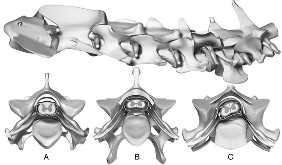
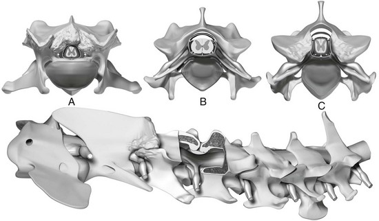
Diagnosis and Treatment of Cervical Spondylomyelopathy

Chapter 236


Diagnosis and Treatment of Cervical Spondylomyelopathy


Cervical spondylomyelopathy (CSM) is a very common disease of the cervical spine of large- and giant-breed dogs. CSM is characterized by dynamic and static compressions of the cervical spinal cord, nerve roots, or both leading to variable degrees of neurologic deficits and neck pain. CSM certainly is a controversial disease! Few other diseases in veterinary medicine have been called by 15 different names, with wobbler syndrome, cervical vertebral instability, cervical malformation/malarticulation syndrome, and disk-associated wobbler syndrome some of the more commonly used. Furthermore, few other disorders have been the target of 27 different proposed surgical treatments. This diversity reflects in part the lack of understanding regarding mechanisms of CSM.


Although the disease can affect essentially all canine breeds, two account for approximately 60% to 70% of all cases: the Doberman pinscher and the Great Dane. These breeds also illustrate the two distinct forms of the disease: disk-associated CSM (primarily affecting Dobermans) and the osseous form of CSM (affecting Great Danes).


Causes and Pathophysiology


The cause of CSM remains unresolved. Proposed causes include genetic, congenital, body conformation, and nutritional factors, with the latter two playing a less significant role. Based on current evidence CSM appears to be congenital. The genetic contribution to the development of the disease remains unclear, although recent evidence in Dobermans suggests that CSM is inherited as an autosomal dominant trait with variable penetrance in this breed.


The pathophysiology of CSM involves both static and dynamic factors. The key static factor is vertebral canal stenosis. It may be an absolute vertebral canal stenosis, which causes direct spinal cord compression and neurologic signs, or a relative vertebral stenosis, which by itself does not lead to myelopathic signs but predisposes the patient to development of myelopathy. Despite some degree of overlap, the spinal cord compressions can be divided into osseous compression and disk-associated compression based on pathophysiology.


Disk-associated compression typically is seen in middle-aged large-breed dogs (mostly Dobermans). It is caused by intervertebral protrusion with or without hypertrophy of the dorsal longitudinal ligament or ligamentum flavum (Figure 236-1). Three factors act in combination to explain the pathophysiology of disk-associated CSM: (1) relative vertebral canal stenosis, (2) more pronounced torsion in the caudal cervical spine leading to intervertebral disk degeneration, and (3) protrusion of larger-volume disks into the caudal cervical spine. Affected dogs apparently are born with a congenital vertebral canal stenosis that predisposes them to the development of clinical signs.


The vast majority of disk-associated spinal cord compressions are located in the caudal cervical spine, affecting the disks at C5-6 and C6-7. The biomechanical features of the caudal cervical spine explain the high incidence of lesions there. This region experiences three times more axial rotation or torsion than the cranial cervical spine, and torsion (more than axial compression) is the main biomechanical force leading to intervertebral disk degeneration in nonchondrodystrophic dogs. Additionally, a study found that Dobermans with CSM have larger intervertebral disks than clinically normal Dobermans (da Costa et al, 2006b). This would cause a larger volume of disk protrusion into the vertebral canal. Since dogs with CSM have a narrow vertebral canal, the combination of a stenotic vertebral canal and protrusion of disks with larger volumes ultimately leads to clinical disease.


The pathophysiology of osseous or bony CSM is different. Osseous CSM is seen predominantly in young adult giant-breed dogs, especially in Great Danes. Although a hereditary basis still is unproven, a familial predisposition has been identified. Giant breeds usually have severe absolute vertebral canal stenosis secondary to proliferation of the vertebral arch (dorsally), articular facets (dorsolaterally), or articular facets and pedicles (laterally) (Figure 236-2). The cause of the compression appears to be a combination of vertebral malformation and osteoarthritic-osteoarthrotic changes at the level of the articular facets. Even though most giant-breed dogs have osseous compression, occasionally these compressions are complicated by disk protrusion, especially in older dogs. Ligamentous compression (by the ligamentum flavum) may be involved in the pathophysiology of the disease in giant- and large-breed dogs, but pure ligamentous compression as the single source of compression appears uncommon.


Critical to understanding the development of clinical signs in CSM-affected large-breed dogs is the concept of the dynamic lesion. Dynamic spinal cord compressions are present in both disk-associated and osseous forms of CSM. A dynamic lesion is one that worsens or improves with changes in the position of the cervical spine. Continuous flexion and extension of the cervical spine can lead to spinal cord elongation causing axial strain and stress within the spinal cord, and this has been proposed as a key mechanism of spinal cord injury in cervical spondylotic myelopathy in humans. This is very different from instability, which has been defined as loss of the ability of the cervical spine to maintain its normal pattern of displacement under physiologic loads and thereby prevent damage to the spinal cord or nerve roots. Instability has not yet been proven in dogs with CSM, and it appears that with the severe disk and osseous degenerative changes, a restricted rather than an excessive motion occurs at the affected sites.


The most common location of compressive lesions in both large- and giant-breed dogs is at C5-6 and C6-7. The lesion is located at one of these sites in 90% of affected large-breed dogs. In giant-breed dogs, the C4-5 site also is commonly affected. Approximately 50% of large-breed dogs have a single site of spinal cord compression, and 50% have two or more sites of similar severity. In giant-breed dogs approximately 20% of dogs have a single site of compression, whereas 80% have multiple compressive lesions. A computed tomographic (CT)–myelographic study identified lesions affecting the T1-T2 and T2 regions in 14% of giant-breed dogs and the C7-T1 region in 22% of all dogs (da Costa et al, 2012). These lesions were not the primary site of compression but were part of the multiple compressions seen in giant-breed dogs. For this reason, it is important to include the cranial thoracic region in imaging studies of dogs suspected of having CSM.


Diagnosis


As indicated earlier, CSM is primarily a disease of large- and giant-breed dogs. It is seen occasionally in small-breed dogs, but it is unclear whether small dogs also have vertebral canal stenosis. The majority of large-breed dogs (weimaraners, dalmatians) and Dobermans with CSM are older than 3 years of age at presentation (mean age, ~7 years). Great Danes and giant breeds (Mastiffs, rottweilers, Bernese and Swiss mountain dogs) with CSM usually are seen at a younger age. The mean age of giant-breed dogs with CSM is 3.8 years, and the disease may be seen in dogs just a few months old.


History and Clinical Signs


A history of chronic progressive signs persisting for several weeks to months is typical. Acute presentations usually are associated with neck pain. Neck pain or cervical hyperesthesia is a common historical finding but is not the typical reason for presentation. Neck pain is part of the clinical findings in approximately 65% to 70% of Dobermans and in 40% to 50% of dogs of other breeds, but it is the chief complaint in only 5% to 10% of dogs with CSM. Forceful manipulations of the cervical spine are unnecessary to document the presence of neck pain and can lead to severe neurologic decompensation. Careful assessment of posture and evaluation of voluntary range of motion (side to side, ventrally and dorsally) using a food treat is recommended to assess for cervical pain. Deep palpation of the transverse processes also can assist in the identification of neck pain.


Gait evaluation is the most important component of the examination in dogs suspected of having CSM because it reliably identifies proprioceptive ataxia, even in the absence of conscious proprioceptive deficits. Proprioceptive ataxia is seen in most dogs with CSM. Dogs with lesions in the cranial or midcervical spine tend to have ataxia affecting all four limbs more uniformly. However, affected dogs typically have obvious pelvic limb ataxia with milder abnormalities in the thoracic limbs. In some cases, the thoracic limb ataxia or weakness may be very mild in comparison with the pelvic limb signs, so that the thoracic limb abnormalities go unnoticed. The thoracic limb gait can appear short-strided or spastic with a pseudohypermetric (“floating”) appearance. Occasionally thoracic limb lameness can be seen, which suggests nerve root entrapment. The pelvic limb gait often is wide based (abducted) and markedly incoordinated. The stride length of the pelvic limbs is increased, which causes the swaying movements of the hind end that are typical of the disease. Scuffing of the pelvic or thoracic limb toes and nails also can be seen. Postural reaction deficits (proprioceptive positioning deficits) are seen in most dogs with CSM but may not be evident in those with a history of long-standing signs despite the presence of proprioceptive ataxia. The reason for this discrepancy is that different tracts carry the pathways for conscious and unconscious proprioception. Approximately 10% of dogs with CSM have nonambulatory tetraparesis at initial presentation.


Evaluation of the spinal reflexes in dogs with CSM indicates a lesion located at either the C1 to C5 spinal cord segments (normal to increased spinal reflexes in all four limbs, with neurologic signs as described earlier) or C6 to C8 spinal cord segments. A C6 to C8 myelopathy is typical because the osseous and disk lesions are concentrated in the C5-6 and C6-7 intervertebral regions. In these cases, the gait is affected in all four limbs but more severely in the pelvic limbs. Evaluation of the spinal reflexes in the thoracic limbs shows a decreased flexor (withdrawal) reflex indicating involvement of the musculocutaneous nerve from the C6 to C8 spinal cord segments, with normal to increased extensor tone suggesting an upper motor neuron lesion and release of the radial nerve from spinal cord segments C7, C8, and T1, mostly C8 and T1. The pelvic limb reflexes are normal to increased.

< div class='tao-gold-member'>

Stay updated, free articles. Join our Telegram channel

Jul 18, 2016 | Posted by in PHARMACOLOGY, TOXICOLOGY & THERAPEUTICS | Comments Off on Diagnosis and Treatment of Cervical Spondylomyelopathy

Full access? Get Clinical Tree

Get Clinical Tree app for offline access