18 Dacryocystitis – chronic purulent conjunctivitis
PRESENTING SIGNS
Dacryocystitis is inflammation within the lacrimal sac and nasolacrimal duct. Affected animals are presented with ocular discharge – usually this is mucopurulent in nature. Alternatively, a unilateral red eye due to conjunctivitis with conjunctival hyperaemia and a variable discharge can be the initial presenting sign. It is quite common for the patient not to be brought to the surgery when the signs first develop, but only when they change and become persistent with a chronic mucopurulent discharge. The discharge is most copious at the medial canthus and reforms frequently following cleansing and removal by the owner. Sometimes the medial canthal skin is erythematous or swollen and the patient is uncomfortable. The condition is rarely encountered in cats but occurs occasionally in dogs and quite frequently in rabbits. The latter are discussed separately in Chapter 19.
CASE HISTORY
The patient may have history of a wet eye 1–3 weeks prior to the development of the chronic discharge and the discharge itself could have changed from serous to purulent in nature. A previous conjunctivitis or even removal of a conjunctival foreign body might have occurred. The patient can be any age or breed. Although normally unilateral in dogs, the condition can be bilateral on occasion (this is more common in rabbits, as discussed in Chapter 19).
CLINICAL EXAMINATION
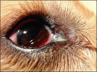
General clinical examination is usually normal in dogs. Ophthalmic examination reveals a unilateral ocular discharge centred around the medial canthus (Figure 18.1). Evidence of pain with blepharospasm or self-trauma though rubbing is variable. Conjunctival hyperaemia is normally present in the nictitans conjunctiva but can be widespread. Schirmer tear test readings are normal. No corneal disease is usually present and the rest of the ocular examination is unremarkable. On cleaning the discharge (after taking a swab for culture and sensitivity testing and cytology for Gram stain) gentle pressure at the medial canthus will often result in the appearance of further purulent material at the nasolacrimal punctae (often just the lower one but sometimes the upper as well). Fluorescein passage to the nostril is absent on the affected side (Figure 18.2); remember to compare both sides, and also that false negatives are common.
CASE WORK-UP
If retrograde cannulation or flushing is not feasible, then further case work-up will involve radiography – both plain and contrast studies (dacryocystorhinography). Plain radiographs might reveal bony fractures or neoplastic processes which were not clinically noted. Periodontal disease involving the upper premolars or canine teeth might be present, leading to a localized inflammation in the nasolacrimal duct and subsequent obstruction. Contrast studies, using water-soluble contrast agents, can locate the exact blockage and help identify whether surgical intervention is feasible. In dogs, small amounts (1–2 ml) of contrast are injected into the upper punctum while holding the lower one closed, such that the contrast material is forced down the duct. Care should be taken to ensure that no excess contrast material is spilled as this could easily confuse interpretation of the radiographs. It is sensible to take a lateral radiograph after injecting the affected side, followed by dorsoventral or intraoral views after injecting both sides for comparison. Cystic dilations can be delineated by this method, as well as locating radiolucent foreign material. If the blockage is anterior – within the lacrimal bone for example – then referral for surgical removal can be considered.
Stay updated, free articles. Join our Telegram channel

Full access? Get Clinical Tree


