20 Cornea – introduction
Diseases of the cornea are frequently encountered in general practice and affect both dogs and cats (as well as rabbits). They can be broadly divided into those involving corneal ulceration and those which are non-ulcerative, with various subdivisions depending on cause, age at onset, appearance and so on (Table 20.1). Specific details of each disease and its management are discussed in the following sections – in this introductory part we will consider various anatomical and physio-logical factors which are applicable to most corneal disease.
Table 20.1 Classification of corneal disease*
| Ulcerative | Non-ulcerative |
|---|---|
* Bold type indicates cases discussed.
ANATOMY/PHYSIOLOGY REFRESHER
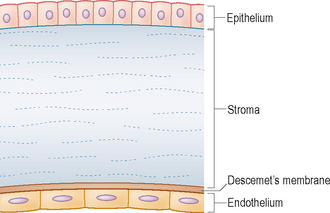
The cornea is the transparent anterior portion of the fibrous outer layer of the globe. It is made up of several layers – the outer epithelium, the middle stroma and the inner endothelium. The acellular layer of Descemet’s membrane lies between the stroma and endothelium (Figure 20.1). In dogs and cats the corneal thickness is approximately 0.45–0.55 mm. The epithelium itself is multi-layered and non-keratinized and produces its basement membrane. Cells are attached to each other and to the basement membrane via interdigitations of the cell membranes and hemidesmosomes. The stroma is the thickest layer of the cornea, made up of multiple layers of fine collagen-containing cells. Descemet’s membrane is an elastic, acellular layer between the stroma and endothelium. The endothelium itself consists of just one layer of cells and therefore is the thinnest cellular layer. Despite its small size, the endothelium is critical for normal corneal functioning as will be seen later on. The corneal surface is bathed by the pre-ocular tear film. Further details of this can be found in the section on the nasolacrimal system (Chapter 15 and cases in Chapters 16 and 17).
Stay updated, free articles. Join our Telegram channel

Full access? Get Clinical Tree


