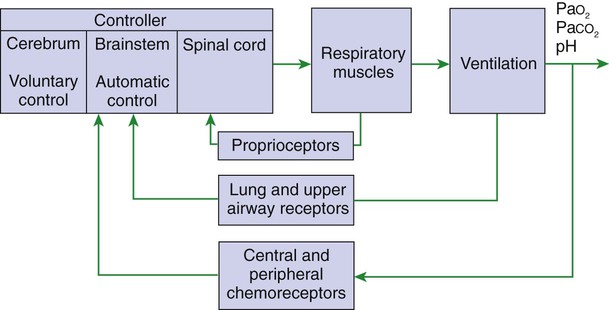
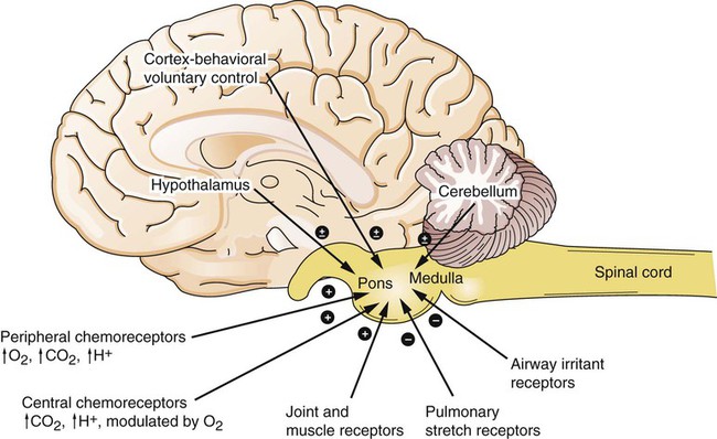
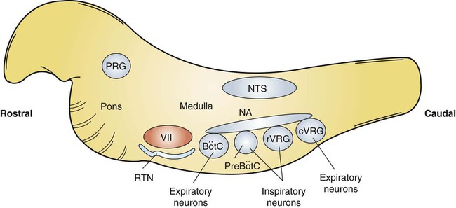
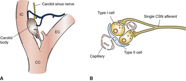
Control of Ventilation


Control of Ventilation


Respiration Is Regulated to Meet the Metabolic Demands for Delivery of Oxygen and Removal of Carbon Dioxide


During its daily activities, an animal varies its level of activity and can breathe air of varying composition and purity. To allow the respiratory system to respond to these different challenges, control mechanisms monitor (1) the chemical composition of the blood, (2) the effort being exerted by the respiratory muscles on the lungs, and (3) the presence of foreign materials in the respiratory tract. This information is integrated with the other nonrespiratory activities, such as thermoregulation, vocalization, parturition, and eructation, to produce a pattern of breathing that maintains gas exchange.


Figure 49-1 provides a diagram of feedback control for the respiratory system. The central controller generates the signals that regulate the activity of the respiratory muscles, which by contracting give rise to alveolar ventilation. Changes in alveolar ventilation affect blood gas tensions and pH, which are monitored by the chemoreceptors. These receptors send signals back to the central controller so that necessary adjustments can be made to ventilation. Mechanoreceptors in various parts of the respiratory system monitor the degree of stretch of the lungs and changes in the airways and vasculature. Stretch receptors (proprioceptors) in respiratory muscles monitor the effort of breathing.


Central Control of Respiration


Respiratory Rhythmicity Originates in the Brainstem with Inputs from Higher Centers


Early attempts to understand the brain’s role in the regulation of breathing indicated that rhythmic respiration originates in the medulla and pons of the brainstem but is also affected by other afferent information from the lungs, chemoreceptors, and elsewhere. After many years of investigation it is becoming clear that the apparently simple, in and out nature of breathing is the result of a complex neural network known as a central pattern generator (CPG) located in the brainstem. This CPG may actually involve rhythmic and nonrhythmic subnetworks that are modulated by inputs from peripheral and central chemoreceptors, pulmonary stretch receptors, and others (Figure 49-2). The status of these subnetworks is affected by sleep, wakefulness, and changes during early development. The CPG includes neurons in the pontine respiratory group (lateral parabrachial and Kolliker-Fuse areas) and several medullary areas especially the Bötzinger and pre-Bötzinger complexes, the retrotrapezoid nucleus (RTN), and the rostral ventral and caudal ventral respiratory groups (rVRG and cVRG, respectively) (Figure 49-3). A dorsal respiratory group of neurons located in the nucleus tractus solitarius relays information to the CPG from peripheral chemoreceptors, pulmonary stretch receptors, bronchial irritant receptors, and other visceral information.


The origin of rhythmic breathing is currently unknown. Pacemaker neurons have been identified in the pre-Bötzinger complex, but their role in normal breathing is unclear. Normal respiration (eupnea) seems to result from reciprocal inhibition of neuron groups in the CPG. During inhalation there is an increase in activity in the inspiratory neurons of the pre-Bötzinger group and rVRG that is associated with diaphragm and external intercostal contraction. This increased activity is further amplified by an increase in the chemical respiratory drive, such as hypoxia or hypercapnia. Termination of inspiration can be a result of vagal inputs from pulmonary stretch receptors or from a central pontine off switch. After vagotomy, the pontine off switch terminates inhalation after a fixed time for inhalation, which is independent of chemical drive. When the vagus is intact, and thus signals from pulmonary stretch receptors are relayed to the brain, there is a complex interaction between the time for inhalation and the tidal volume. This interaction leads to a larger tidal volume and more rapid respiratory frequency when the chemical drive to breathe is increased.


When inhalation is terminated, inspiratory neurons are inhibited, and thus exhalation occurs passively as a result of the elastic recoil of the lung and chest wall. Activity in some inspiratory neurons (Bötzinger complex) early in exhalation leads to inspiratory muscle activity, which provides a “brake” on exhalation and regulates the rate of expiratory airflow. Later in exhalation, the braking is removed. During this latter part of exhalation, expiratory neurons may be activated (Bötzinger complex and cVRG) leading to expiratory (abdominal and internal intercostal) muscle contraction. When respiratory drive is low, this second phase of exhalation is initiated later than when drive is increased.


The rhythmic breathing just described is frequently overridden by demands from higher brain centers. Vocalization, parturition, swallowing, defecation, and many other activities require the active participation of the respiratory system.


Pulmonary and Airway Receptors


Pulmonary Stretch Receptors, Irritant Receptors, and Juxtacapillary Receptors Can Influence the Rhythm of Breathing


The vagus nerve includes both myelinated and nonmyelinated afferent axons conveying sensory information from the lung. Myelinated axons originate from slowly adapting stretch receptors and irritant receptors. Slowly adapting stretch receptors are nerve endings associated with smooth muscle in the trachea and main bronchi, but to a lesser degree in the smaller intrapulmonary airways. They are stimulated by deformation of the wall of larger airways, as when intrathoracic airways are stretched during lung inflation. Because firing rates from these receptors increase progressively as the lung inflates, they are thought to be responsible for the inhibition of breathing caused by lung inflation (Hering-Breuer reflex). Termination of input from these receptors by vagotomy leads to a slowing of respiration and an increase in tidal volume. Slowly adapting stretch receptors may be responsible in part for adjustments in the rate and depth of respiration to minimize the work of the respiratory muscles.


Irritant receptors, or rapidly adapting stretch receptors, are thought to be myelinated nerve endings branching among epithelial cells in the larynx, trachea, large bronchi, and intrapulmonary airways. They are stimulated by mechanical deformation of the airways, such as the deformation that occurs during mechanical irritation of the airway surface. Irritant gases, dusts, mucus accumulations, histamine release, and a variety of other stimuli can also cause these receptors to respond. Stimulation of rapidly adapting irritant receptors leads to cough, bronchoconstriction, mucus secretion, and rapid, shallow breathing (hyperpnea), all of which are protective responses to clear irritant materials from the respiratory system. These receptors may initiate the sighs that are thought to redistribute pulmonary surfactant over the alveolar surface.


C fibers are located in the pulmonary interstitium close to pulmonary capillaries (juxtacapillary receptors), where they may monitor blood composition or the degree of distention of the interstitium. Similar fibers also occur in the walls of the airways. C-fiber activation may be responsible for the increase in respiratory rate (tachypnea) that accompanies allergic, infectious, or vascular diseases.


In addition to intrapulmonary receptors, there are receptors located in the upper airway. Stimulation of receptors in the nasal cavity causes sniffing and sneezing, whereas stimulation of laryngeal and pharyngeal receptors may cause cough, apnea, or bronchoconstriction. Temperature receptors in the pharynx that are cooled by airflow alert the animal if there is insufficient airflow so that appropriate adjustments can be made by the inspiratory muscles to increase flow.


Chemoreceptors


Hypoxia, Acidosis, and Hypercapnia Are All Potent Stimuli for Ventilation


Chemoreceptors monitor oxygen, carbon dioxide, and hydrogen ion concentration (pH) at several sites in the body and provide some tonic drive to respiration during normal breathing. As blood composition departs from normal, changes in arterial carbon dioxide (PaCO2) and oxygen (PaO2) tensions and pH produce major changes in ventilation.


Peripheral Chemoreceptors Are the Only Receptors Monitoring Blood Oxygen Levels but Also Respond to Changes in Carbon Dioxide and Hydrogen Ion Concentrations


Peripheral chemoreceptors include the carotid and aortic bodies, and their removal eliminates the respiratory response to hypoxia. The response to CO2 and pH persists because these are also detected by central chemoreceptors.


The carotid bodies (Figure 49-4, A) are located close to the bifurcation of the internal and external carotid arteries, and the aortic bodies are located around the aortic arch. The latter appear to be most active in the fetus and of little importance in the adult. The aortic bodies are supplied by the vagus nerve, and the carotid bodies are supplied by a branch of the glossopharyngeal nerve. Fibers within the nerves supplying the peripheral chemoreceptors are primarily brainstem afferents, with a few parasympathetic and sympathetic efferent fibers to blood vessels.


< div class='tao-gold-member'>

Stay updated, free articles. Join our Telegram channel

Jul 18, 2016 | Posted by in PHARMACOLOGY, TOXICOLOGY & THERAPEUTICS | Comments Off on Control of Ventilation

Full access? Get Clinical Tree

Get Clinical Tree app for offline access