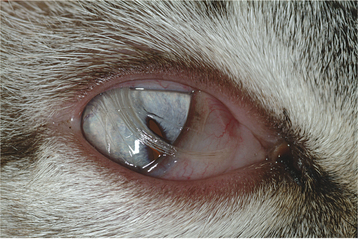
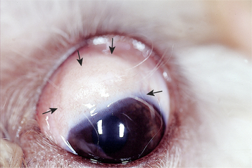
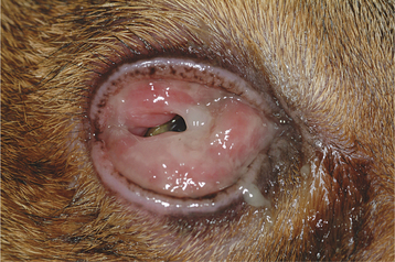
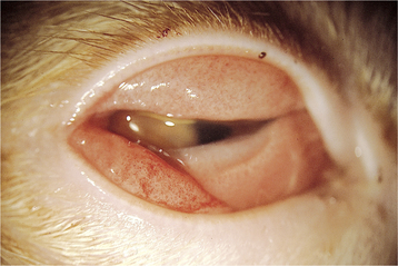
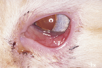
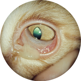
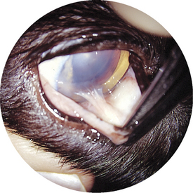
Chapter 2 Conjunctiva INTRODUCTION The conjunctiva is a highly vascular layer on the surface of the eye and eyelids. It functions in some respects as a lymph node does, reacting to antigens to which the ocular surface is exposed. The most frequently diagnosed condition is conjunctivitis, which may have various causes (e.g., infectious, allergic). Infectious conjunctivitis and its sequelae are seen commonly in cats. Conjunctival lesions in dogs and cats, including neoplasia, are illustrated in this chapter. Figure 2-1 Dermoid arising from the dorsal conjunctiva in a 5-month-old Siamese cat. A dermoid is an occurrence of normal, haired skin in an abnormal location. Dermoids are found on eyelid margins, conjunctiva, and corneas. Figure 2-2 Large conjunctival dermoid in a young dog. Most of the dermoid is not haired. Figure 2-3 Severe herpesvirus conjunctivitis in a cat. Note the marked conjunctival swelling (chemosis) and pseudodiphtheritic membrane formation. Figure 2-4 Conjunctivitis with chemosis and petechia in a cat with herpesvirus infection. Figure 2-5 Conjunctivitis associated with feline herpesvirus infection in an adult cat. Chemosis, conjunctival injection, and mild mucopurulent ocular discharge are present. Figure 2-6 Conjunctivitis associated with feline herpesvirus infection in a cat. Upper respiratory tract infection, chemosis, conjunctival injection, and mucopurulent ocular discharge are present. Figure 2-7 Symblepharon caused by a resolved herpesvirus infection in a cat. A symblepharon is an adhesion between structures of the eye; in this case an adhesion exists between the bulbar surface of the nictitans and the cornea.< div class='tao-gold-member'> Only gold members can continue reading. Log In or Register to continue Share this: Share on X (Opens in new window) X Share on Facebook (Opens in new window) Facebook Related Related posts: Lacrimal System Orbit Glaucoma Eyelid Stay updated, free articles. Join our Telegram channel Join Tags: Color Atlas of Canine and Feline Ophthalmology Jul 31, 2016 | Posted by admin in INTERNAL MEDICINE | Comments Off on Conjunctiva Full access? Get Clinical Tree
Chapter 2 Conjunctiva INTRODUCTION The conjunctiva is a highly vascular layer on the surface of the eye and eyelids. It functions in some respects as a lymph node does, reacting to antigens to which the ocular surface is exposed. The most frequently diagnosed condition is conjunctivitis, which may have various causes (e.g., infectious, allergic). Infectious conjunctivitis and its sequelae are seen commonly in cats. Conjunctival lesions in dogs and cats, including neoplasia, are illustrated in this chapter. Figure 2-1 Dermoid arising from the dorsal conjunctiva in a 5-month-old Siamese cat. A dermoid is an occurrence of normal, haired skin in an abnormal location. Dermoids are found on eyelid margins, conjunctiva, and corneas. Figure 2-2 Large conjunctival dermoid in a young dog. Most of the dermoid is not haired. Figure 2-3 Severe herpesvirus conjunctivitis in a cat. Note the marked conjunctival swelling (chemosis) and pseudodiphtheritic membrane formation. Figure 2-4 Conjunctivitis with chemosis and petechia in a cat with herpesvirus infection. Figure 2-5 Conjunctivitis associated with feline herpesvirus infection in an adult cat. Chemosis, conjunctival injection, and mild mucopurulent ocular discharge are present. Figure 2-6 Conjunctivitis associated with feline herpesvirus infection in a cat. Upper respiratory tract infection, chemosis, conjunctival injection, and mucopurulent ocular discharge are present. Figure 2-7 Symblepharon caused by a resolved herpesvirus infection in a cat. A symblepharon is an adhesion between structures of the eye; in this case an adhesion exists between the bulbar surface of the nictitans and the cornea.< div class='tao-gold-member'> Only gold members can continue reading. Log In or Register to continue Share this: Share on X (Opens in new window) X Share on Facebook (Opens in new window) Facebook Related Related posts: Lacrimal System Orbit Glaucoma Eyelid Stay updated, free articles. Join our Telegram channel Join