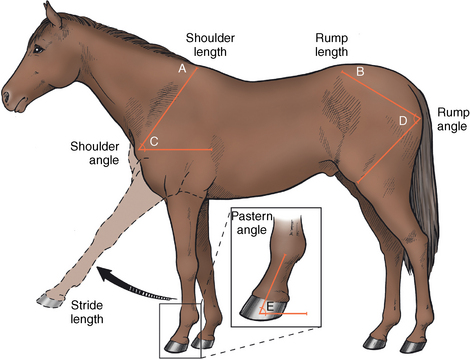
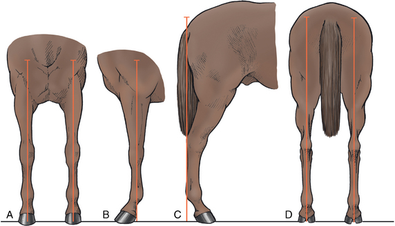
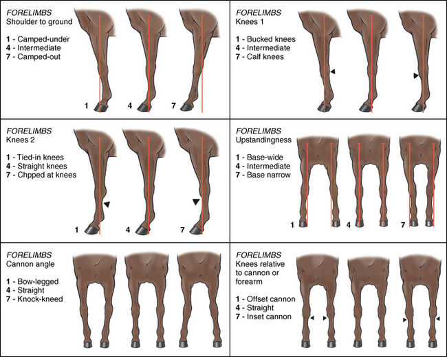
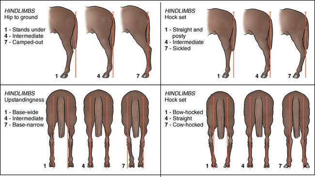
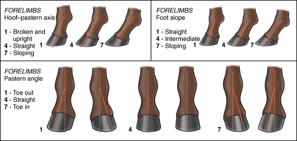
CHAPTER 15 Conformation can be defined as the “formation of something by appropriate arrangement of parts or elements: an assembling into a whole” (Webster’s dictionary, 1976) and equine conformation appraisal is traditionally based on the external appearance of the body shape, form or outline of the animal. This evaluation may be regarded as the front line for judgments when selecting horses for specific intended tasks, including breeding selection. Prepurchase recommendations and perceived animal value rest highly on this assessment. There is wide variation of conformation between and within different breeds, the significance of which requires expert understanding of optimal breed characteristics and potential effects on soundness or performance. The success of a horse in any equine discipline or industry is not dependent on “perfect” conformation, as this does not guarantee performance or soundness, and “imperfect” conformation does not necessarily exclude a horse from performing at elite levels. Other factors such as human management, environmental conditions, genetics, nutrition, temperament, training, and the health status of the horse will also have a large bearing on ultimate performance. Conformation can, therefore, only be considered an indicator for future athletic potential. Nonetheless, conformation can assist prediction of possible musculoskeletal strengths and weaknesses, possible predisposition to injury, or both, based on known etiology and pathophysiology of musculoskeletal disorders. The conformation or inherent anatomic structure of the horse is an integral part of the equine musculoskeletal constitution and will influence the quality of dynamic performance. The skeletal format will affect such factors as joint range of motion, limb arc and hoof flight patterns, and weight distribution in motion, with subsequent effects on coordination of movement (including limb interference), balance, power (propulsion, impulsion, and collection), agility, and endurance. Conformation will, therefore, partially dictate the relationship between form and function, thus modifying the potential for biomechanical efficiency, superior performance, musculoskeletal durability, and perhaps even longevity (Wallin et al., 2001). As some conformational traits are dynamic and will only be apparent during ambulation, the traditional emphasis of conformational assessment as a pure description of static external appearance has been extended to include a more functional assessment of conformation during unridden and ridden gaits in some of the studies cited in this chapter. There is a great need to clarify and standardize the descriptive terminology of joint alignments, as most conformational traits are described using multiple traditional and variable nonscientific terms, rather than by defining anatomic configuration. For example, a caudal deviation at the radiocarpal or metacarpal joint complex (knee) may be described as “back at the knee,” “calf knee,” or “carpal hyperextension,” none of which describes the precise origin of segmental misalignment. Biomechanical evaluation relies heavily on strict physical and mechanical relationships of segments, requiring accurate anatomic terminology. Yet, most studies have employed generalized or horsemanship terms in describing conformational traits. The lack of anatomic precision, documentation, or both limits the interpretation of some studies. The literature presented in this chapter will follow the terminology appearing in the research papers. Some common terms describing conformational alignments are defined anatomically in Table 15-1 and illustrated in Figure 15-1 and Figure 15-2. TABLE 15–1 Anatomic Description of Commonly Used Conformational Terms FIGURE 15-1 Illustrations of some common conformational defects of the forelimbs (see Table 15-1 for description). (From Mawdsley A, Kelly EP, Smith FH, Brophy PO: Linear assessment of the thoroughbred horse: an approach to conformation evaluation, Equine Vet J 28:461, 1996.) FIGURE 15-2 Illustrations of some common conformational defects of the hindlimbs (see Table 15-1 for description). (From Mawdsley A, Kelly EP, Smith FH, Brophy PO: Linear assessment of the thoroughbred horse: an approach to conformation evaluation, Equine Vet J 28:461, 1996.) All assessment of equine conformation should be conducted with the horse standing squarely (loading all limbs symmetrically) on a level surface. The stance of the horse has been identified as a major source of error in conformation assessment, as small changes in limb placement and weight distribution can introduce significant variation in segmental alignment. When assessing deviation of the limb from the vertical, Weller et al. (2006a) found measurement variations in stance within one horse to be almost as large as between horses, thus highlighting the importance of standardized repeatable positioning of the horse. Conformation assessment should be a systematic and organized process incorporating a general overall observation of size, symmetry, musculature, posture, balance, and demeanor, followed by a more specific evaluation of conformational traits of the body, individual limbs, and feet. Briefly, relevant body observations should include head shape and size; height at the withers and croup; body length; neck length; shoulder length (top of the withers to point of the shoulder); pelvic length (tuber coxae to tuber ischii); scapular and humeral inclination; pelvic and femoral inclination; and chest width. From these observations, an overall proportioned symmetry in lengths and heights is desirable, both left to right and fore to hind. Congruent sloping angulation of the shoulder and hip is also desirable, with a proportional length of individual limbs in relation to the height and size of the body (Figure 15-3). The segment lengths of specific long bones of limbs should also be noted at this time. Establishing the exact source of the alignment deviation is imperative; for example, does a laterally pointing hoof, commonly described as toed out, originate from an externally rotated limb or from a particular distal joint? Cranial, caudal, and lateral views are needed to determine limb deviations in the sagittal, coronal (frontal), and transverse planes (see Figures 15-1 and 15-2). When examining the conformational traits of individual limbs, a plumb line approach is useful in identifying angular or torsional deviation of segments from the vertical or horizontal at each joint level (Figure 15-4). In horses with “ideal” conformation, a visualized vertical plumb line dropped from the tuberosity of the scapular spine should bisect the longitudinal axis of the forelimb to the metacarpophalangeal joint (MCPJ or fetlock) and fall 5 cm behind the heel in the lateral view. A line dropped from the cranial aspect of the greater tubercle of the humerus (point of the shoulder) should bisect the forelimb in the cranial view. In the hindlimb, a plumb line dropped from the ischial tuberosity should touch the point of the calcaneous (prominent caudally in the tarsus or hock), follow the plantar metatarsal surface to the metatarsophalangeal joint (MTPJ or fetlock), and fall 7.5 to 10 cm (Ross, 2003) caudal to the heel in the lateral view. The entire hindlimb should be bisected evenly in the caudal view (see Figure 15-4). When assessing foal conformation, limbs can also be viewed from above at the shoulder and hip (skyline view). Particular attention is warranted in evaluation of distal limb alignment, hoof quality, size, and balance due to the concentration of locomotive stresses in this area. Although different breeds will have feet of different shapes and sizes, it is universally and anecdotally desirable to have balanced feet positioned symmetrically under the central limb axis with a straight hoof–pastern axis (the dorsal surface of the hoof wall lies parallel to the dorsal surface of the pastern region) (see Figure 15-3 and Figure 15-5). The constant growth of the hoof creates a dynamic relationship between the digital axis and dorsal hoof wall, which suggests that completely straight hoof–pastern axes cannot exist over time without natural wear or appropriate trimming (Moleman et al., 2006). FIGURE 15-5 Illustrations of some common conformational defects of the hooves (see Table 15-1 for description). (From Mawdsley A, Kelly EP, Smith FH, Brophy PO: Linear assessment of the thoroughbred horse: an approach to conformation evaluation, Equine Vet J 28:461, 1996). After assessment, overall observations can be related to desirable or “benchmark” breed-specific conformational characteristics and judgment made on the horse’s suitability to a given career. Notably, the definition and number of traits evaluated, the point scale scoring system of conformational traits, and the image of an ideal phenotype varies greatly among registries, organizations, and countries; therefore, specific classification is essential for comparative evaluations. Selection of a horse in the presence of a less-than-desirable conformation is not always considered unwise. A study on Thoroughbred racehorses highlighted that variation in horses and performance is not fully explained by a few underlying conformational components but is a result of a complex interaction of all conformational parameters (Weller et al., 2006b). However, certain conformational faults such as extreme tarsal angulation (large or small) and tarsal valgus are almost certainly predisposing to injury or lameness in racing events and are best avoided. Veterinarian conformational assessment should particularly focus on the presence of any such faults and the relationship of these faults to existing or potential pathologic conditions (Rossdale and Butterfield, 2006). In many instances, coexisting conformational anomalies will be present, at times allowing biomechanical compensation and at other times exacerbating musculoskeletal stresses during locomotion. The evaluation of conformation has traditionally been subjective or empirical and remains the primary method of assessment. Visual appraisal of defined criteria (the outlines and axes described above) and manual palpation of specific bony landmarks have been the basis of assessment, giving the examiner multiple three-dimensional images over a period. The combinations of joint configurations and segment lengths are infinite and multifaceted, so the resulting judgment is variable and directly dependent on the individual expertise and personal “ideal” of the practitioner. Magnusson (1985) showed less variance among judges on overall impressions and type traits. However, opinions concerning segment lengths, joint angles, and skeletal inclinations were largely discrepant. This finding was supported by a study comparing radiographic and visual assessments of hoof–pastern conformation in Warmblood foals (Kroekenstoel et al., 2006).Visual assessment was only in agreement with radiologic evidence in 6 of 92 (6.5%) evaluations. Weller et al. (2006c) also suggested that variability in judgment is affected by the limited repeatability of measurement techniques due to inaccurate identification of anatomic landmarks and inconsistent positioning of the subject. Some studies and studbooks have used a system of linear scoring in an attempt to quantify the repeatability of subjective evaluation (Dolvik and Klemetsdal, 1999; Koenen et al., 1995; Mawdsley et al., 1996). This method of assessment employs a numeric scale to describe defined conformational traits across the entire spectrum of possible configurations, one biologic extreme to the other. Although meeting with some success, 6 of 21 traits were classified unacceptably low in repeatability (Mawdsley et al., 1996). These traits were hoof–pastern axis in both forelimbs and hindlimbs, head size, and vertical alignment of the forelimbs and hindlimbs, all having a coefficient of variation greater than 10%. Despite these limitations, subjective evaluation can be easily and quickly performed by an experienced evaluator, expediting the assessment of large numbers of horses within a short time frame. The absence of standardized evaluation standards, lack of centralized training programs internationally, and a large source of error introduced by subjective assessment precludes sole use of this method to compare results between studies or substantiate the more complex relationships among conformation, performance, and soundness. For these, quantitative conformational assessment, in addition to these traditional judging methods, has been suggested to improve predictive capability (Holmstrom and Philipsson, 1993). Initial attempts to provide absolute values in conformation assessment have used the tools listed in Table 15-2 in combination with a reference marker system. A founding study by Magnussen (1985) described the comprehensive set of landmarks listed below, and many research studies have followed this protocol or a derivative of it. TABLE 15–2 Tools of Conformation Measurement 1. Cranial end of the wing of atlas 2. Proximal end of the spine of the scapula 3. Caudal part of the greater tubercle 4. Transition between the proximal and the middle thirds of the lateral collateral ligament of the elbow 5. Lateral tuberosity of the distal end of the radius 6. Space between the fourth carpal, the third metacarpal, and the fourth metacarpal bones 7. Proximal attachment of the lateral collateral ligament of the fetlock joint to the distal end of the third metacarpal bone 1. Proximal end of the tuber coxae 2. Center of the anterior part of the greater trochanter of the femur 3. Proximal attachment of the lateral collateral ligament of the stifle joint to the femur 4. Attachment of the long lateral ligament of the tibiotarsal joint to the plantar border of the calcaneus 5. Space between the fourth tarsal, the third metatarsal, and the fourth metatarsal bones 6. Proximal attachment of the lateral collateral ligament of the fetlock joint to the distal end of the third metatarsal bone The major disadvantages in using these methods are the possible errors introduced by marker placement on skeletal landmarks, particularly in the proximal skeleton, the consequent reliability of findings, and the time required to perform the measurements (Weller et al., 2006a). Radiography has also been used to measure joint angles and segment lengths. However, this requires expensive equipment, has the health and safety implications of possible radiation exposure to personnel involved, and is very sensitive to subject positioning (Barr, 1994; White et al., 2008). Advancing technology has allowed more objective, quantitative evaluation of conformation amenable to statistical analysis and aims to find evidence-based relationships among conformation, performance, and soundness. This has resulted in verification of some traditional empirical ideals and refuting of others, though results are often conflicting. For global advancement in this area of study, it is clearly imperative to use universally comparative methodology, which is somewhat lacking. Objective conformational evaluation provides a useful adjunct to subjective assessment by quantification of some conformational traits; however, it must be remembered that not all conformational aspects can be measured objectively. Aesthetic factors such as athletic elegance, suppleness, overall balance and harmony, jumping style, and movement symmetry are necessarily subjectively based.
Conformation
Common Term
Anatomic Description
Base narrow
Distance between the forelimbs is greater at the chest than feet, the limb sloping medially
Back at the knee/calf knee
Carpal hyperextension due to a caudal displacement of the proximal row of carpal bones, the radiocarpal joint being <180 degrees (Ross, 2003). An upright pastern is often also related to this conformation (Ducro et al., 2009a)
Forward at the knee/bucked knee/over at the knee/sprung knee
Radiocarpal joint angle >180 degrees or lack of full carpal extension causing a flexion moment
Offset knee/bench knee
Traditionally described as the metacarpus laterally deviated relative to the carpus; however, the displacement is usually in the radiocarpal joint (Ross, 2003)
In at the knee/knock knee
Carpal valgus
Tied in below the knee
Distinct notch distal to the accessory carpal bone on the palmar aspect of the limb causing the circumference of the leg below the carpus to be less than that above the metacarpophalangeal joint (fetlock)
Upright pastern
Metacarpophalangeal and proximal interphalangeal (pastern) joints have a straight appearance
Toed out feet
Metacarpophalangeal valgus
Toed in feet
Metacarpophalangeal varus
Uneven feet
Forefeet differ in size, shape, or both, causing variable hoof–ground angles
Sickle hock/curby hock
Tibiotarsal (hock) angle 53 degrees or less (Holmstrom et al., 1990)
Straight behind
Tibiotarsal angle >170 degrees (Marks, 2000), usually due to a more upright tibia
Cow hocked/in at the hock
Either a rotational change in the hindlimb or tarsus valgus >180 degrees
Assessment of conformation
Subjective assessment of conformation
Objective assessment of conformation
Tool
Measurements Taken
Goniometer (see Figure 15-3)
Joint angles
Scapular/pelvic inclinations
Tape measure
Height at withers
Length of croup and back
Width of chest and mandible
Circumference of girth; neck at poll and withers (Mawdsley et al., 1996); carpus; the third metacarpal/metatarsal; girth
Box level +/– crossbar
Height at withers, back, and croup
Length of head, body, limbs
Depth of chest
Width of breast and pelvis
Calipers
Width of head and third metacarpal/metatarsal
Width of chest and pelvis
Neck and forelimb
Hindlimb
![]()
Stay updated, free articles. Join our Telegram channel

Full access? Get Clinical Tree


Conformation
Only gold members can continue reading. Log In or Register to continue
