Complicated corneal ulcer

23 Complicated corneal ulcer


The previous two case descriptions illustrate very com-mon presentations in general practice – the superficial non-healing ulcer and the deep ulcer. The treatment plan outlines ways to treat these conditions but assumes that everything goes well. In this chapter we will discuss the ulcer that does not heal according to plan.


CLINICAL EXAMINATION


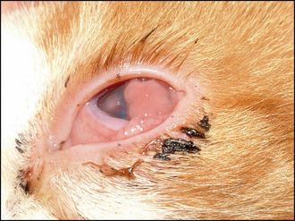
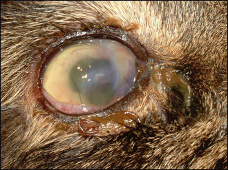
The ulcer should be obvious on clinical examination. However, the conjunctiva is likely to be hyperaemic, the nictitans membrane elevated and chemosis might also be present which could make visualization of the cornea difficult (Figure 23.1). Sometimes the ocular discharge will be extensive such that the eye is ‘glued’ shut. Gently bathing this away (and taking swabs for culture and sensitivity at the same time – but see below) will assist in examination. Overt ulceration might not initially be visible – there is likely to be significant vascularization (even granulation tissue) on the cornea, along with oedema, both of which obscure the actual ulcer. If areas of cornea appear gelatinous, and creamy or grey in colour, this could suggest keratomalacia or corneal melting (Figure 23.2).


To make it easier to examine a painful cornea, the patient should be placed at the end of the consulting table and made to look down, while you look up from below. This usually makes it possible to view most of the cornea as the nictitans membrane moves into the ventromedial fornix. The use of topical anaesthesia, and even sedation or general anaesthesia, might be necessary to fully evaluate the eye in some patients.


CASE WORK-UP


It is essential that swabs are taken for culture and sensitivity testing from all potentially infected corneal ulcers. These can be taken before topical anaesthesia. Samples can be collected from the ocular discharge but more accurate isolation will be obtained from the ulcer edge itself – a bacterial swab moistened with sterile saline can be gently rolled onto the edge of the ulcer. This can also be used to perform Gram staining (by then rolling the swab onto a microscope slide and staining it appropriately). Cytology should also be considered – samples collected by swabs do not yield high numbers of cells and so the use of a cytobrush or Kimura spatula (or the blunt end of a scalpel blade) is preferred. Topical anaesthesia is necessary before such sampling. Samples should be spread onto a microscope slide and air dried. Staining with Diff-Quick or similar stains can be useful to look for the presence of bacteria, the type and number of cells (usually a combination of neutrophils and degenerate keratocytes in a bacterial infection) and the presence of inclusion bodies (for example, with Chlamydophila). The presence of fungal elements should be considered – although relatively rare in the UK, these can have devastating consequences if not identified and treated accordingly.


The eye should be fully examined for the evidence of anything that could be contributing to the poor healing, or exacerbating the ulcer. Retained foreign bodies and abnormal cilia are probably the most common culprits after bacterial infection.


NURSING ASPECTS


The specific nursing for complicated corneal ulcers is similar to those for deep ulcers (see Chapter 22). Thus regular bathing of any discharge and the frequent application of topical medication are necessary. It is also important to alert the attending veterinary surgeon to any change in the condition of the patient, both of the eye itself and of the animal’s general demeanour – a sudden deterioration could require urgent surgical intervention.


EPIDEMIOLOGY


The complication of bacterial infection is the most common cause of ulcer progression. The conjunctival sac normally carries a mixed population of mainly Gram-positive bacteria (e.g. staphylococci, streptococci and corynebacteria). These can act as opportunist pathogens once the corneal epithelium is breached. However, infection with more pathogenic organisms, mainly Gram-negative bacteria (e.g. Escherichia coli, Pseudomonas), is likely in more severe ulcers. Mycotic infections do occur occasionally in the UK but are common in some parts of the world. Cytology is important for their diagnosis initially, followed by culture to identify the exact fungus. Candida albicans and Aspergillus spp. are reportedly the most frequent isolates in the UK.


Not all complicated ulcers are infected. Sometimes degenerating keratocytes or neutrophils can release collagenases which cause further corneal damage. Clearly if other pathological processes are occurring in the eye simultaneously – uveitis or glaucoma for example – the risk of complications with an ulcer is higher.


TREATMENT OPTIONS – MANAGEMENT


The owner should be fully apprised of the severity of the condition from the outset. A treatment plan should be drawn up and followed. Obviously this must be tailored to the individual and will include factors such as patient compliance, owner finances and commitment, as well as the condition of the eye itself.


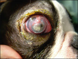
The first decision to be made should be: Is the eye salvageable? In some cases the cornea will be so severely compromised that the ulcer has ruptured, the iris has prolapsed and a panophthalmitis has resulted (Figure 23.3). If this is the case, then enucleation might be the best option for the patient. However, this is an irreversible decision and so one should be convinced that there is no alternative. If the eye has the potential for vision, enucleation is not advised. A positive dazzle response, together with a consensual pupillary light reflex (i.e. constriction of the fellow eye when a bright light is shone into the affected eye), are encouraging signs. If neither is present, then removing the eye might be indicated. If you are not sure whether the eye can be saved, referral should be considered. The welfare of the patient is paramount.


Stay updated, free articles. Join our Telegram channel

Sep 10, 2016 | Posted by in SMALL ANIMAL | Comments Off on Complicated corneal ulcer

Full access? Get Clinical Tree

Get Clinical Tree app for offline access