Chronic epiphora

17 Chronic epiphora


CLINICAL EXAMINATION


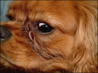
General clinical examination is typically unremarkable. On ophthalmic examination the obvious abnormality is a wet face – tear overflow from the medial canthus runs down the side of the nose (Figure 17.1). Often there is dark staining (as the presumed lactoferrin-type pigments in tears react with the atmosphere), and some localized skin inflammation and moist dermatitis can be present. It is essential to determine whether the tear overflow is purely due to poor drainage (i.e. epiphora) or to increased lacrimation. Sometimes a combination of both can be present. However, if Schirmer tear test readings are normal and there is no evidence of ocular discomfort, then epiphora can usually be assumed.


Careful examination with magnification and a good light source are required to evaluate potential causes for the epiphora. The nasolacrimal punctae should be evaluated for presence, size and position. Any medial entropion should be assessed together with lid apposition – some toy breeds have very tight skin–globe apposition which functionally reduces drainage. The caruncles in the medial canthi should be examined to see if any hairs are present on them which could be wicking tears away from the punctae and down the face. Other facial hair can contact the tear film and cause wicking as well – the fine curling fur of poodles is an important contributor to tear overflow.


Examination for conditions which could cause irritation and increase tear production slightly should be undertaken. Thus the presence of nasal folds rubbing on the cornea or conjunctiva, distichia or ectopic cilia, overt entropion or corneal ulcers should be noted (Figure 17.2). In most cases of pure epiphora these latter abnormalities are not present and the rest of the ophthalmic examination is normal.


image

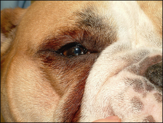
Figure 17.2 Epiphora and increased lacrimation in a young English bulldog. In addition to the tear staining at the medial canthus this dog is wet around the lateral canthus (unlike Figure 17.1) and some lower lid entropion is present causing increased tear production rather than the dog just having poor drainage. Schirmer tear test readings would be high in this dog, but normal in the CKCS in Figure 17.1.


EPIDEMIOLOGY


Once again, as is often the case for ophthalmic conditions, the selective breeding of pedigree dogs has led to the clinical problems encountered. Imperforate or micropunctae can occur in any breed, but golden retrievers and cocker spaniels (both English and American) are certainly over-represented, together perhaps with the Cavalier King Charles spaniel, miniature and toy poodle, Samoyed and Bedlington terrier.


The tear overflow syndrome seen in the miniature and toy poodle, Maltese terrier and so on is partly due to the head shape and ocular appearance of these dogs which has been specifically chosen by breeders. The small, tight eyelids can lead to medial entropion and physical closure of the punctae which are frequently already small. Unfortunately, the same problems are now seen in brachycephalic cats as well, where selection for the short nose and prominent eyes has led to medial entropion, tight medial lid–globe apposition and trichiasis in exactly the same way as in some dogs. Since in many patients the epiphora does not cause any clinical problem to the animal, and is therefore purely cosmetic, breeders do not see that this is still an abnormality which further selective breeding could reduce significantly.


Sep 10, 2016 | Posted by in SMALL ANIMAL | Comments Off on Chronic epiphora

Full access? Get Clinical Tree

Get Clinical Tree app for offline access