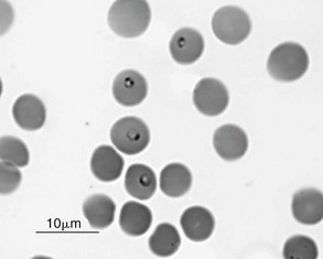
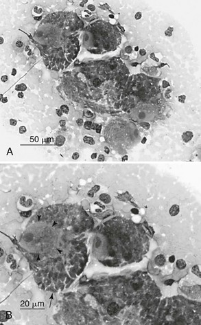
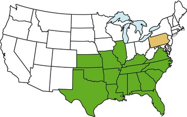
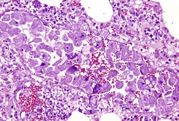
Web Chapter 84 Cytauxzoon felis is a tick-transmitted intracellular protozoon of emerging importance that infects wild and domestic cats. In domestic cats it can cause severe disease characterized by hemolytic anemia and circulatory impairment that is associated with high mortality. Historically, C. felis infection was considered a uniformly fatal disease in cats; however, recently several reports have described cats surviving infection. The genus Cytauxzoon belongs to the subphylum Apicomplexa, order Piroplasmida, and family Theileriidae, which are parasites of mammals. Cytauxzoon organisms exist in two distinct tissue forms: an erythrocyte phase (piroplasm) (Web Figure 84-1) and a tissue phase (schizont) (Web Figure 84-2). The genus name Cytauxzoon is used to designate species reported initially in African ungulates in which the tissue phase develops in mononuclear phagocytes lining the blood vessels of numerous organs, whereas species in the genus Theileria invade lymphocytes during the schizogonous phase. Cytauxzoon is related closely to the genus Babesia; however, Babesia organisms have exclusively an erythrocyte stage without a tissue phase. Web Figure 84-1 Peripheral blood from a cat with cytauxzoonosis. Four piroplasms are present within erythrocytes. Two of the piroplasms (top) each contain two nuclear areas. Two of the erythrocytes (bottom) contain small particulate stain precipitate that must be differentiated from organisms. (Wright-Giemsa stain.) (From Meinkoth J, Kocan AA: Feline cytauxzoonosis, Vet Clin North Am Small Anim Pract 35:89, 2005, with permission.) Web Figure 84-2 A, Impression smear of spleen from a cat with cytauxzoonosis. Five large schizont-containing macrophages are present. Scattered myeloid cells and plasma cells are also seen. B, Higher magnification of the area shown in A. The nucleus of a host cell is outlined by arrowheads. The host cell nucleus contains a greatly enlarged nucleolus. The schizont completely fills the cell’s cytoplasm, often being indistinct. In portions of the cell multiple merozoites are seen forming within the schizont, giving the cytoplasm a “packeted” appearance (arrow). (Wright-Giemsa stain.) (From Meinkoth J, Kocan AA: Feline cytauxzoonosis, Vet Clin North Am Small Anim Pract 35:89, 2005, with permission.) C. felis was first recognized in 1976 in southwestern Missouri. It now has been reported in the south central, southeastern, mid-Atlantic, and Gulf Coast states of the United States (Web Figure 84-3). The pattern of geographic distribution probably is affected by the availability of both reservoir hosts (wild bobcats) and arthropod vectors (Dermacentor variabilis, Amblyomma americanum). The natural life cycle of C. felis is not completely understood. The domestic cat was presumed to be an accidental terminal host in which C. felis caused predominantly a rapidly fatal disease, but recent findings of asymptomatic infected cats have led to the possibility that domestic cats serve as an additional reservoir for C. felis. In contrast, bobcats (Lynx rufus) are the presumed reservoir hosts acting as persistent carriers of the organism and usually developing only mild or subclinical infection. There is a paucity of information on blood parasites of free-ranging cats, however, and Cytauxzoon has been reported in bobcats only since 1982, so questions remain as to whether this is a new disease of felids or only a newly identified disease. Mountain lions (Felis concolor) from Florida and Texas also have been reported to carry the organism. There has been a correlation of disease with tick interchange among wild and domestic felids, with ticks presumably transmitting the organism between cats by feeding. In addition to D. variabilis (American dog tick), A. americanum (Lone Star tick) recently has been demonstrated experimentally to be a competent vector for Cytauxzoon. Both tick species are common in the southeastern and midwestern United States where the majority of C. felis infections have been identified. Transstadial transmission occurs in ticks but transovarial transmission has not been demonstrated. However, cytauxzoonosis may be more widespread than previously recognized because cytauxzoonosis-like diseases have been reported in domestic cats in Zimbabwe and Spain, in Pallas’s cats (Otocolobus manul) in Mongolia, and in a lion (Panthera leo) in Brazil. Experimental studies have revealed no zoonotic or agricultural risk for C. felis. Web Figure 84-3 Map of the United States showing states in which there have been confirmed cases of Cytauxzoon felis infection in either domestic cats (green) or bobcats (gold). (From Cohn LA: Cytauxzoon infections. In August JR, editor: Consultations in feline internal medicine, ed 6, St Louis, 2010, Saunders, with permission.) After infection, the organism undergoes schizogony, an asexual reproductive phase, in mononuclear phagocytic cells associated with vessels of almost every organ. As the schizonts (see Web Figure 84-2) undergo schizogony and binary fission, macrophages lining the blood vessels enlarge tremendously and occlude venules in liver, spleen, lung (Web Figure 84-4), and lymph nodes, which result in a thrombuslike mechanical obstruction of blood flow and tissue hypoxia. The schizont is associated with clinical disease, and the greater the schizont burden the more severe the clinical illness. Infected domestic cats have extensive schizont burdens, whereas mildly affected bobcats have a limited and transient schizogonous phase. The tissue phase also is suspected to release toxic, pyrogenic, and vasoactive products that contribute to clinical manifestations. The schizogonous tissue phase, detectable by 12 days after infection, is responsible for the venous congestion, thrombotic disease, and organ failure that leads to death in most cats within 3 weeks of infection. Web Figure 84-4 Lung tissue with pulmonary venule from a cat with cytauxzoonosis showing near-total occlusion of the vessel by large macrophages parasitized by merozoites within the cytoplasm. Developing organisms give a granular appearance to the cytoplasm. (Hematoxylin and eosin stain.) (From Snider TA, Confer AW, Payton ME: Pulmonary histopathology of Cytauxzoon felis infections in the cat, Vet Pathol 47:698, 2010, with permission.)
Feline Cytauxzoonosis


Epidemiology

Pathogenesis

![]()
Stay updated, free articles. Join our Telegram channel

Full access? Get Clinical Tree


