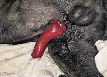
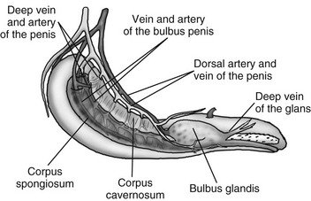
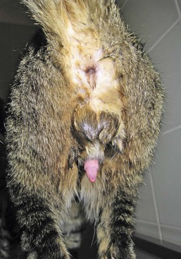
Web Chapter 73 Priapism is a persistent penile erection lasting longer than 4 hours without sexual stimulation. Priapism easily can be confused with paraphimosis. Paraphimosis occurs when the nonerect penis cannot be ensheathed in the prepuce and can result secondarily in edema. However, the penis is not erect as with priapism. Paraphimosis can result from inadequate length of the prepuce, weakened preputial muscles, too small of a preputial orifice, or trauma (Web Figures 73-1 and 73-2). Web Figure 73-2 Paraphimosis in an 11-month-old Cane Corso mastiff. The penis is swollen markedly and cannot be ensheathed in the prepuce. The canine erection is mediated through the pelvic nerve, a parasympathetic nerve, arising primarily from the first and second sacral nerves (S1-S2). Stimulation of the pelvic nerve dilates penile arteries, partially inhibits venous drainage, and increases penile blood pressure, causing an erection. Initially blood flow to cavernous bodies increases, as the muscles in the helicine branches of the deep arteries and the arteries of the urethral bulb relax. The corpus spongiosum receives a larger share of blood from the artery of the bulb of the penis. Arterial blood then is shunted to the corpus cavernosum from the artery of the bulb (Web Figure 73-3). The deep vein of the penis becomes unable to drain the increased arterial blood flow into the cavernous spaces from the helicine arteries. Internal pressure against the tunica albuginea results in the corpus cavernosum becoming stiff. The intrinsic veins subsequently become compressed. A second stage occurs after intromission when partial venous occlusion is a factor in the erection of the glans penis. Contraction of the female’s constrictor vestibulae muscles stimulates reflexive contraction of the male’s ischiourethralis muscle. Arterial flow increases and is directed into the dorsal artery of the penis. The pudendal nerve, arising from S1-S3, also is involved by stimulating contraction of the extrinsic penile muscles. Ischiourethralis muscle contraction decreases venous blood flow through the dorsal veins. Venous blood is shunted from the corpus cavernosum toward the bulbus glandis. The relatively thick muscular-walled arteries prevent arterial flow from being occluded by the extrinsic muscle contraction. Venous connections with the corpus spongiosum and valves in the deep veins of the glans prevent blood from exiting the bulbus except through the currently inefficient dorsal penile veins. Relaxation of the intersinusoidal trabecular smooth muscles facilitates pars longa glandis and bulbus glandis engorgement, increasing penile stiffness. The hypogastric nerve, a sympathetic nerve that originates from L1-L4, also may have a regulatory role in the canine erection. Hypogastric nerve stimulation in the dog causes increased blood flow into the cavernous space secondary to vasodilation of the inflow blood vessels. However, an inhibitory effect also may occur because of relaxation of the outflow blood vessels, increasing cavernous blood outflow. The hypogastric nerve is responsible for prostatic secretion and ejaculation. Web Figure 73-3 Anatomy and vascular supply of the canine penis. (Illustration courtesy Alex Frederick.)
Priapism in Dogs

Physiology of Erection

Chapter 73: Priapism in Dogs
Only gold members can continue reading. Log In or Register to continue

Full access? Get Clinical Tree


 -year-old Savannah cat.