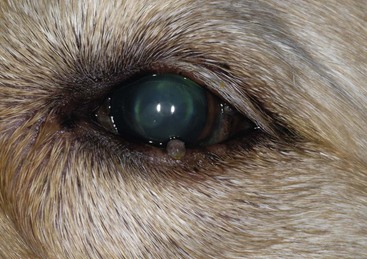
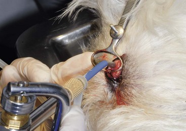
Canine Ocular Neoplasia

Chapter 257


Canine Ocular Neoplasia


Ocular neoplasia is relatively uncommon compared with neoplasia affecting other body systems; however, it is the second most common cause of enucleation. Early detection of ophthalmic neoplasia is important for optimal treatment outcome. Ophthalmic disease, particularly uveitis, can be the first sign of systemic neoplasia, and multicentric neoplasia can involve the eye, which makes an ocular examination desirable in all canine oncology patients. These relationships emphasize the importance of a complete physical examination in all cases of canine uveitis. Additionally, owners may be less willing to pursue oncologic treatment in blind pets, which underscores the importance of early detection and aggressive treatment of ocular neoplasia.


General Diagnostic Approach


In general, a working diagnosis should be made based on ophthalmic and physical examination findings and results of fine-needle aspiration (FNA) or biopsy. Appropriate staging tests can be selected based on these results. For orbital tumors, computed tomography (CT) and magnetic resonance imaging (MRI) are very helpful in determining the local extent of disease and planning surgical and radiation treatment. For tumors involving the globe, ocular ultrasonography is useful for identifying and localizing masses obscured by corneal, aqueous, or lens opacity and for those located in the posterior segment. Ocular ultrasonography also is useful for guiding FNA, and high-resolution ultrasonography can be used to assess the extent of tumors in the cornea, sclera, and iris. Thoracic radiography and lymph node aspiration should be performed to detect metastatic disease, with recognition that the overall metastatic rate for most malignant ocular neoplasms is low. The eye also may be a site of metastatic or multicentric neoplasia (as in the case of lymphoma), so the initial diagnosis will influence the subsequent diagnostic and treatment decisions.


For eyelid lesions, which typically are benign, early diagnosis is the key to complete resolution without the need for complicated surgical reconstruction. One third of the eyelid margin length can be excised without impairing eyelid function. As for other skin lesions, cytologic analysis, examination of skin scrapings, and FNA are straightforward diagnostic procedures for periocular masses. The eye must be protected or retropulsed whenever a needle or blade is directed toward the globe, so chemical restraint often is indicated to prevent ocular trauma during the procedure.


For conjunctival lesions, most diagnostic procedures can be performed using topical anesthesia (proparacaine hydrochloride 0.5% solution) and gentle manual restraint. A sample of a conjunctival lesion for cytologic analysis can be obtained with the blunt end of a scalpel blade, a cytobrush, or a Kimura spatula. Biopsy of the conjunctiva also can be performed using topical anesthesia. The conjunctiva adjacent to the lesion should be grasped using small, toothed forceps and gently elevated, and then the lesion should be snipped free with small scissors. The sample then can be gently spread onto a tongue depressor for fixation. Alternatively, the sample can be placed directly into a histopathologic tissue cassette. Closure of conjunctival defects smaller than 4 mm in diameter is unnecessary. Although biopsy may be more invasive, it provides more information than cytologic analysis alone and may prove essential to obtaining a diagnosis. Subconjunctival or episcleral masses most often are inflammatory, but differentiating these from a neoplastic mass can be a diagnostic challenge. Biopsy of an episcleral mass may warrant referral to an ophthalmologist to avoid inadvertent penetration of the globe.


Examination of the third eyelid also can be performed with topical anesthesia. Nontoothed forceps may be used to grasp the leading margin and elevate the third eyelid, which allows the palpebral and bulbar surfaces to be visualized and palpated directly. Biopsy of the third eyelid conjunctiva may be performed as described earlier. For larger lesions for which excisional biopsy or removal of the entire third eyelid may be warranted, general anesthesia may be required. Because the development of keratoconjunctivitis sicca is a possible complication of complete third eyelid excision, it is important to ensure that informed consent is obtained before this procedure is performed.


Aqueous paracentesis is performed infrequently on visual eyes because it requires general anesthesia; carries a (low) risk of bacterial endophthalmitis, profuse iridal hemorrhage, or retinal detachment; and reliably induces transient uveitis. Aqueous humor cytologic evaluation is most sensitive for the diagnosis of lymphoma, if the diagnosis cannot be made by sampling other tissues. Vitreocentesis is most useful in cases of exudative or solid posterior segment masses in blind globes. For blind eyes, aqueous humor cytologic analysis or vitreal aspirate examination may yield a diagnosis before enucleation that may improve surgical planning, but enucleation is preferred in nonvisual eyes affected with neoplasia.


For orbital masses, FNA, collection of culture specimens, and Tru-Cut biopsy can be performed through the conjunctiva, periocular skin, or oral cavity behind the last molar. Tru-Cut biopsy is best performed under sedation or anesthesia in conjunction with ultrasonography or CT to avoid damage to the globe. Plain radiographs are useful mostly for identifying dental or nasal disease or osteolysis; CT or MRI is far superior for evaluating the soft tissues of the orbit and globe as well as for assessing involvement of orbital bone and sinuses. Orbital mucoceles and abscesses tend to have a more cavitary appearance than orbital tumors on ultrasonographic, CT, or MRI scans.


Signalment, history, and examination findings help further with differential diagnoses. For instance, dental disease and neoplasia are more common causes of orbital disease in older patients. Abscesses tend to arise more quickly and to elicit more pain than tumors. Application of topical lubricants or performance of a temporary tarsorrhaphy often is needed in cases of severe exophthalmos to protect the cornea while a definitive diagnosis is being made and therapy is implemented.


Histopathologic diagnosis of ophthalmic neoplasia can be complicated by the idiosyncrasies of ophthalmic anatomy and tumor physiology, species differences, the mount of inflammation present within a tumor, the orientation of the tissue, and the experience of the veterinary pathologist. Submitting a detailed history as well as drawings and photographs of the lesion in situ, inking the margins of adnexal tumors, and marking the scleral location of intraocular tumors with suture after enucleation are recommended to assist the pathologist in making the most accurate and informative diagnosis. When an enucleated globe is submitted for histopathologic analysis, fixation in a volume of formalin 10 times that of the globe is optimal for adequate fixation of intraocular tissue. A 25-gauge needle may be used to inject approximately 0.5 ml of formalin through the sclera posterior to the equator of the globe to improve fixation of the intraocular structures.


Primary Ocular Neoplasia


Adnexa and Conjunctiva


The most common canine eyelid neoplasms are meibomian gland adenoma and epithelioma (Figure 257-1) These tumors are benign but locally expansive, arising from the sebaceous glands and glandular epithelium, respectively. They typically occur in older dogs, have an irregularly textured to cobblestone surface, and can be seen protruding from the meibomian gland orifice as a pink, gray, or black mass at the eyelid margin. If the tumor remains within the meibomian gland it may cause obstruction of the gland orifice, resulting in accumulation of the glandular lipid secretions within the duct. When rupture of the gland ensues, marked granulomatous inflammation may develop in response to the release of inflammatory lipid products into the surrounding tissues. This secondary inflammation may give a small meibomian gland adenoma a falsely large appearance and can be quite uncomfortable for the patient. Meibomian gland adenomas and epitheliomas also can cause significant corneal irritation, and although these rarely are a direct cause of corneal ulceration, they may prevent epithelialization and delay corneal wound healing when an ulcer is present.


Squamous papillomas, which arise from the eyelids and periocular skin, have a clinical appearance and behavior similar to those of tumors of meibomian gland origin and also are frequent in older dogs. Squamous papillomas in younger dogs are more likely of viral origin and usually regress spontaneously without any treatment. Benign eyelid melanomas also are common in older dogs and typically are broad-based pigmented masses adjacent to but not usually involving the eyelid margin. Pedunculated forms rarely occur.


Surgical resection is the treatment of choice for most canine eyelid tumors. Surgical options include full-thickness eyelid resection, carbon dioxide laser ablation, and surgical debulking with cryoablation. When full-thickness eyelid resection is performed, it is imperative to achieve perfect reapposition of the eyelid margin to ensure normal eyelid function and long-term corneal health. Surgical debulking with cryoablation is an alternative to full-thickness eyelid resection that does not require general anesthesia in most patients. It can be performed under sedation (e.g., dexmedetomidine 375 to 500 µg/m2 IV and butorphanol 0.1 mg/kg IV) and local anesthesia (0.5 ml of lidocaine 2% infiltrated into the base of the neoplasm). After an open-closed chalazion clamp is placed over the eyelid and tumor, small scissors are used to excise all neoplastic tissue visibly extruding from the meibomian gland orifice. Gentle digital pressure may be applied to the palpebral conjunctiva to extrude any glandular contents. If the tumor can be visualized through the palpebral conjunctiva, a No. 15 Bard-Parker scalpel blade can be used to sharply excise the overlying conjunctiva to the level of the tumor, which allows débridement with a 3- or 4-mm curette or sharp excision using Stevens tenotomy scissors. It is important not to incise the eyelid margin using this debulking approach and to ensure that any incisions extend only through the palpebral conjunctiva. After the majority of the tumor material has been removed, cryoablation can be performed using liquid nitrogen in a dispensing canister with a flat probe attached (Figure 257-2). The size of the ice ball generated provides an estimate of the depth of cryopenetration. Use of two complete freeze-thaw cycles is recommended. The size of the tip selected for the cryoablation unit should approximate the size of the base of the tumor. Postoperatively, patients should be treated for 5 to 7 days with a broad-spectrum ophthalmic antibiotic ointment. Swelling and epidermal depigmentation occur commonly at the site of cryoablation, but repigmentation may be expected within weeks to months. In rare cases depigmentation remains at the eyelid margin, which may be a concern in patients for which cosmesis is important.


< div class='tao-gold-member'>

Stay updated, free articles. Join our Telegram channel

Jul 18, 2016 | Posted by in PHARMACOLOGY, TOXICOLOGY & THERAPEUTICS | Comments Off on Canine Ocular Neoplasia

Full access? Get Clinical Tree

Get Clinical Tree app for offline access